Clear Sky Science · en
Multimodal AI-based 28-day mortality prediction of pneumonia patients at ED discharge: a multicenter study
Why this study matters for everyday patients
Pneumonia is one of the world’s deadliest infections, yet in a busy emergency department doctors often must decide within minutes whether a patient is safe to go home or needs intensive hospital care. This study explores whether artificial intelligence (AI) can combine chest X‑ray images with routine medical data to more accurately predict who is at high risk of dying within 28 days, helping clinicians avoid sending fragile patients home or admitting low‑risk patients unnecessarily.
How pneumonia care works today
When someone arrives at the emergency department with suspected pneumonia, clinicians rely on symptoms, vital signs, blood tests, and a chest X‑ray to judge how sick the person is. For years they have used simple point‑based scores such as CURB‑65, which add up factors like age, blood pressure, and confusion to estimate the chance of death over the next month. These scores are easy to calculate but ignore much of the information now routinely collected, and they do not use the wealth of detail hidden in chest X‑ray images. As a result, some patients who later deteriorate may initially look “low risk” on paper.
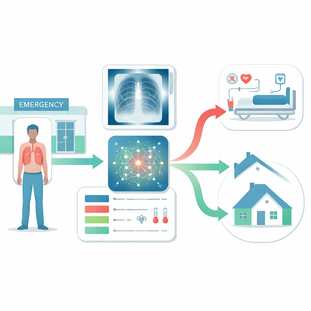
Bringing images and data together
Recent advances in deep learning allow computers to read chest X‑rays and quantify findings such as fluid around the lungs or suspicious spots. The researchers in this study asked whether feeding those computer‑generated image findings into a more sophisticated prediction model, together with standard emergency department data, could sharpen short‑term risk estimates. They drew on records from nearly 2,900 emergency visits for pneumonia at a major hospital in South Korea and from the large MIMIC‑IV database in the United States. For each visit, they collected three main types of information: the traditional CURB‑65 elements, the AI’s numerical interpretation of the chest X‑ray, and automatically captured clinical data such as vital signs, oxygen needs, and routine laboratory results taken before the patient left the emergency department.
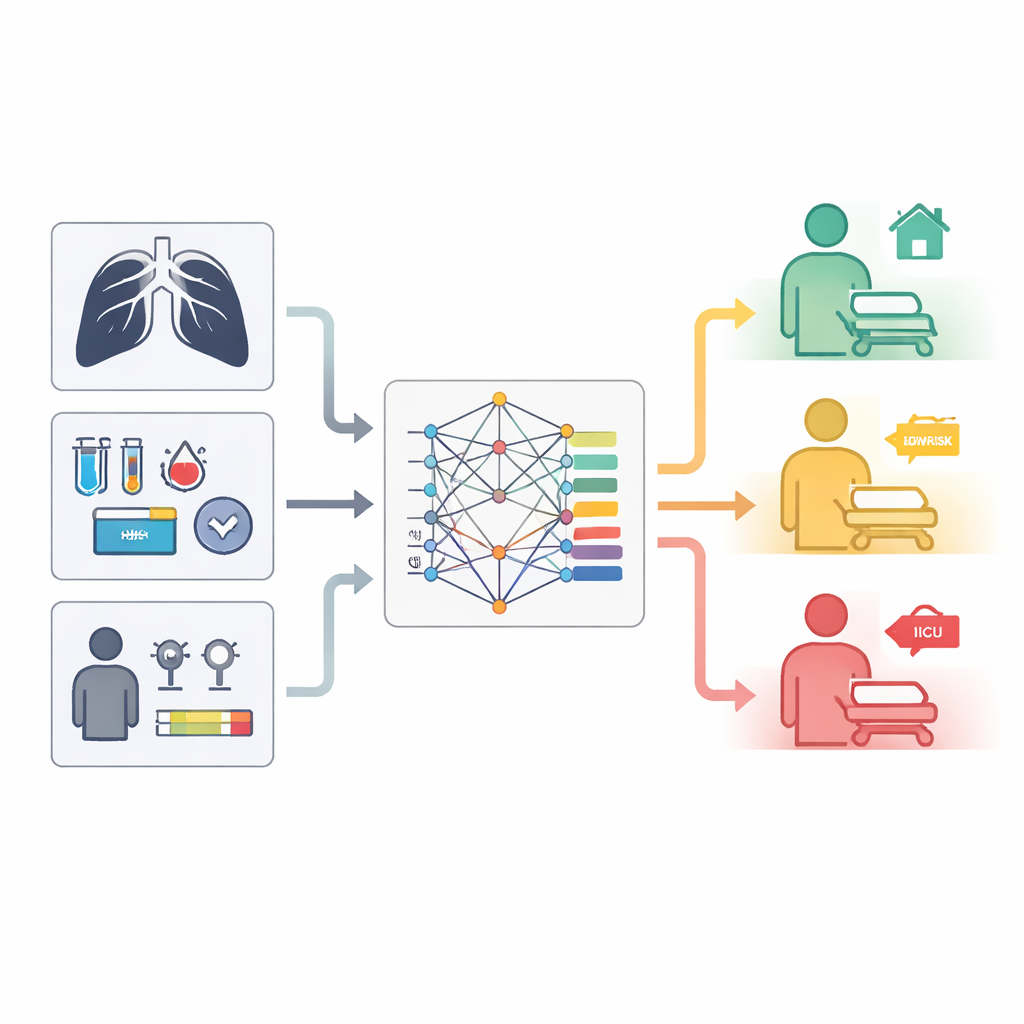
Building and testing the AI model
The team compared several approaches, including a conventional statistical model and modern machine‑learning methods designed to handle time‑to‑event outcomes. Their most powerful approach, called a random survival forest, was trained to learn complex patterns linking the combined data to the chance of death within 28 days. They created versions of this model using different input sets: only the traditional score, only image findings, only clinical variables, mixtures of score plus clinical data, and finally an “all‑features” model that included everything. Performance was judged mainly by how well each model could correctly rank patients from lowest to highest risk, a measure known as the concordance index.
What the model learned about risk
The model that used all available information—traditional score components, AI‑interpreted chest X‑ray findings, and detailed clinical data—performed best. It correctly ordered patients by risk substantially more often than the CURB‑65 score alone. Even compared with a model that used rich clinical data but ignored image information, adding the chest X‑ray output gave a measurable improvement. Age and triage acuity emerged as the strongest predictors, aligning with clinical experience that older, sicker patients fare worse. Yet image features such as the likelihood of fluid around the lungs and small nodules also ranked among the most influential factors, showing that the AI was extracting useful signals from X‑rays that standard scores do not capture. When the researchers grouped patients into “high” and “low” risk, their full model identified nearly all of the patients who died within 28 days, while keeping the number of false alarms similar to that of the traditional score.
Designed to fit into real hospital workflows
Importantly, the system was built to work with information that hospitals already collect automatically. It drew on demographics, vital signs, oxygen support level, and laboratory values from the electronic record, plus the output of a chest X‑ray AI tool that was already in routine use. The researchers deliberately excluded elements that demand extra human judgement or manual data entry, such as bedside assessments of confusion, to avoid adding to clinicians’ workload. They also used explanation techniques to check that the model’s behavior made clinical sense, and they iteratively refined which variables were included based on both mathematical importance and expert review. This mix of machine insight and human oversight is meant to increase trust and ease adoption at the bedside.
What this could mean for patients and hospitals
The study suggests that an AI model that merges chest X‑ray interpretations with routine emergency department data can more accurately flag pneumonia patients at high risk of dying within a month than long‑standing scoring tools. In practice, such a system could help emergency teams prioritize scarce hospital beds for those who need them most, ensure timely treatment for vulnerable patients who might otherwise be sent home, and safely reduce admissions for those at genuinely low risk. While the work still needs broader testing in other hospitals and with different imaging software, it points toward a future in which quietly running algorithms support, rather than replace, clinicians by turning everyday data into sharper, more individualized risk estimates.
Citation: Hwang, S., Heo, S., Hong, S. et al. Multimodal AI-based 28-day mortality prediction of pneumonia patients at ED discharge: a multicenter study. Sci Rep 16, 11761 (2026). https://doi.org/10.1038/s41598-026-42378-2
Keywords: pneumonia, emergency department, artificial intelligence, chest x-ray, risk prediction