Clear Sky Science · en
The first limb-sparing use of histotripsy for canine osteosarcoma
Saving a Leg Without a Knife
Bone cancer in large-breed dogs is not only common, it is often heartbreaking: the usual way to control the tumor is to amputate the affected limb. This study explores a different path. Instead of surgery, the researchers tested a new ultrasound-based approach called histotripsy to break up bone tumors while leaving the limb in place. Their work in pet dogs living with naturally occurring cancer hints at a future where both dogs and people with similar tumors might keep better mobility, experience less pain, and still receive powerful local treatment.
A Tough Cancer Shared by Dogs and People
Osteosarcoma is the most common primary bone cancer in both dogs and humans, and it behaves aggressively in each. Many patients eventually die from spread of the disease to the lungs or other sites, even after major surgery and chemotherapy. In dogs, amputation or complex limb-salvage operations are standard, but these procedures can lead to infection, hardware failure, repeat surgeries, and long recoveries. Because large-breed dogs develop osteosarcoma far more often than humans and progress through the disease more quickly, they offer a powerful real-world model for testing new treatments that could help both species.
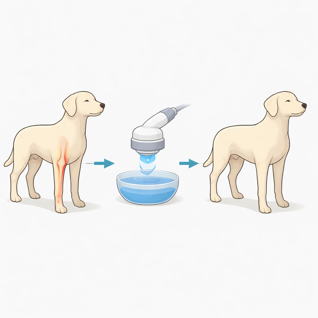
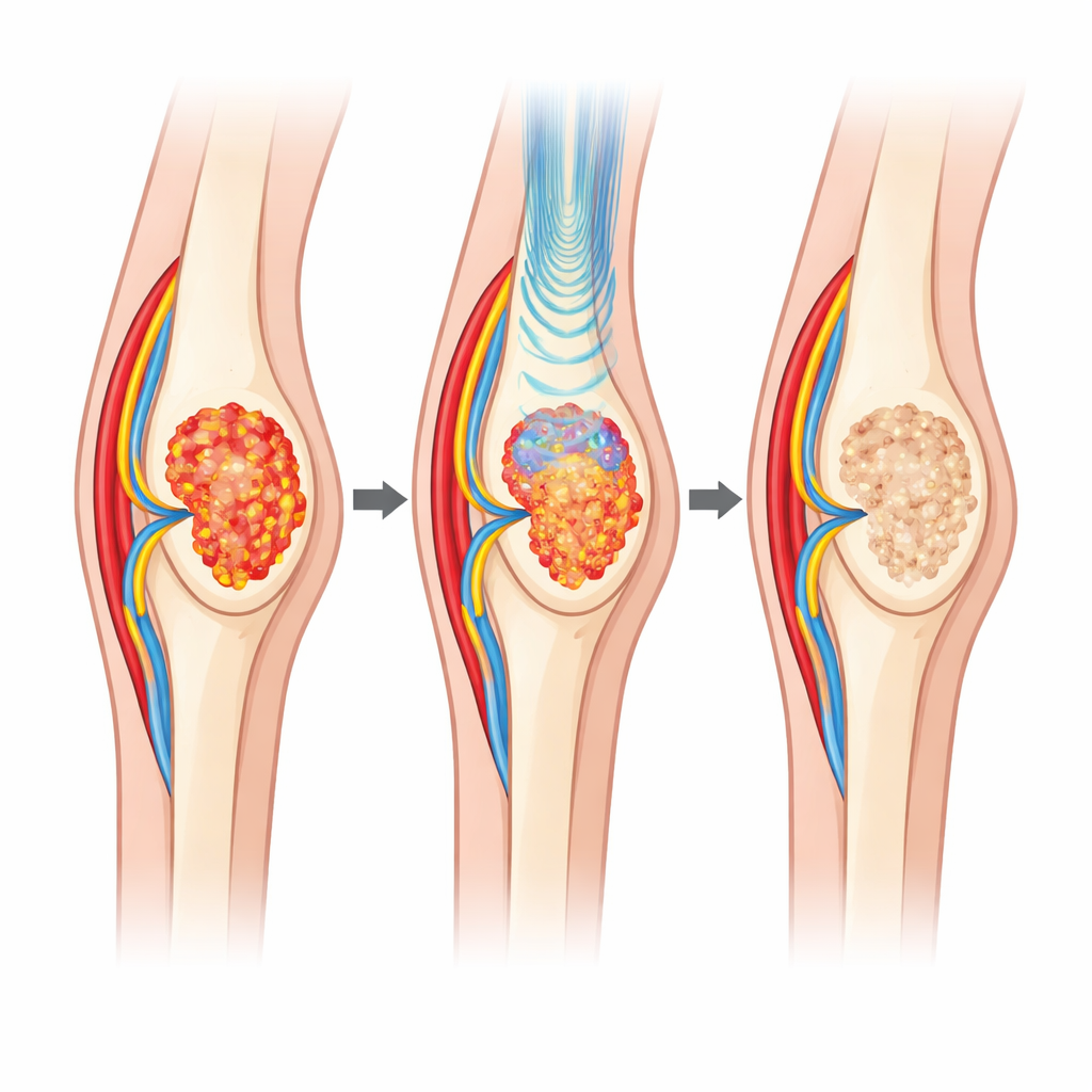
A Gentle Hammer Made of Sound
Histotripsy uses precisely focused ultrasound pulses to create tiny bubbles inside tissue. These bubbles rapidly expand and collapse, mechanically shredding targeted cells into a fine slurry while sparing nearby structures that are mechanically tougher, such as major blood vessels, nerves, and healthy bone. Unlike heat-based ultrasound treatments, histotripsy does not rely on cooking tissue, which reduces the risk of burns and damage outside the target. Early trials in canine bone cancer had only treated small parts of tumors that were later removed by amputation. In this new work, the team went further: they attempted to destroy large portions of bone tumors, over several sessions, without taking the limb off afterward.
How the Trial Worked in Real Pets
Nine pet dogs with suspected limb bone tumors were enrolled after their owners declined standard options like amputation, radiation, or chemotherapy. Under general anesthesia, each dog’s tumor was mapped in detail with MRI scans, and a robotic arm guided the histotripsy transducer to focus sound deep within the tumor. Depending on tumor size, each dog received one to five treatment volumes, spaced over several days or weeks, to cover as much of the mass as possible. The team used follow-up MRI scans to see how much of the tumor stopped taking up contrast dye, a sign that it was no longer well supplied with blood and likely no longer viable. They also repeatedly measured how the dogs walked on a pressure-sensing walkway and collected owner surveys about pain and quality of life.
What Changed Inside the Bone and in Daily Life
Imaging showed that histotripsy could carve out well-defined non-enhancing regions within the tumors, often covering more than half of the cancer volume and, in some dogs, effectively all of it. Overall, tumors tended to look slightly larger after treatment—probably from swelling—but their contrast uptake dropped by more than half, implying that much of the tissue inside had been destroyed or devitalized. Four of six dogs with follow-up imaging later developed new active areas mainly at the tumor edges, suggesting that future treatments may need to include a small margin of nearby tissue or be combined with other therapies. Importantly for comfort and function, dogs placed significantly more pressure on their affected limb when walking at the end of follow-up than they did before treatment, a pattern usually linked to less pain. About two-thirds of dogs with available data showed clinically meaningful improvements in at least one pain measure, and on average there was no worsening of lameness, pain, or quality of life.
Risks, Limits, and Hints of What Comes Next
The treatments were generally well tolerated. Out of 24 treatment sessions, four caused moderate to severe skin or soft-tissue injury on the far side of the limb, likely due to heat buildup where there was little tissue to absorb or carry away excess energy. Adjusting the pulse pattern and adding a cooling pad reduced the rate of these problems in later dogs. While the study was small and not designed to prove a survival benefit, some dogs lived and remained free of distant spread longer than typically expected without surgery, and one dog with near-complete tumor ablation had only modest local growth over more than five months. The authors suggest that future trials should test faster ways to ablate large bone tumors, refine protection of skin and soft tissue, and explore whether histotripsy can also stimulate the immune system when paired with modern immunotherapies.
What This Could Mean for Dogs and People
To a non-specialist, the key message is that powerful sound waves might someday replace the scalpel for many bone cancer patients. In this first limb-sparing trial in pet dogs, histotripsy was able to destroy large parts of bone tumors, reduce their blood supply, and often ease pain, while allowing the animals to keep their legs. The procedure did carry some risk of skin injury and did not cure the disease, but it showed that noninvasive, repeatable ultrasound treatments can be delivered safely to stubborn bone tumors. With larger studies and continued refinement, the same approach may eventually offer both canine and human patients a better balance between tumor control and quality of life.
Citation: Vickers, E.R., Ruger, L.N., Hay, A.N. et al. The first limb-sparing use of histotripsy for canine osteosarcoma. Sci Rep 16, 14574 (2026). https://doi.org/10.1038/s41598-026-42319-z
Keywords: osteosarcoma, canine cancer, focused ultrasound, limb-sparing therapy, histotripsy