Clear Sky Science · en
Multicenter ex vivo evaluation of an α-mannosidase–targeted fluorescent probe for intraoperative margin assessment in breast cancer
Why this matters for people facing breast surgery
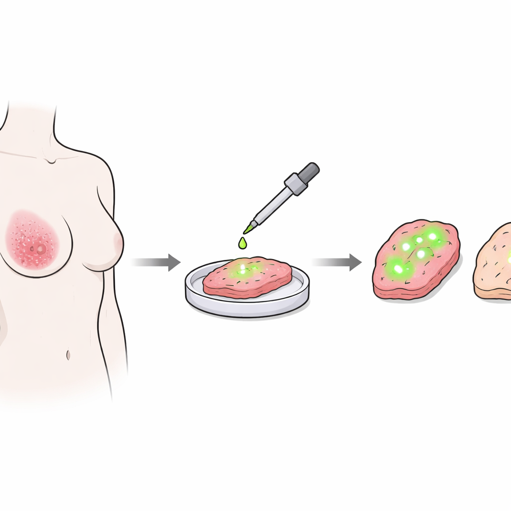
When surgeons remove a breast tumor, they must be sure that no cancer cells are left at the cut edge of the tissue. Today, this often requires time‑consuming lab work during the operation and the judgment of an expert pathologist. This study reports a new glow‑in‑the‑dark style test that can quickly show surgeons whether the removed tissue still contains cancer at its margins, potentially reducing repeat surgeries and stress for patients.
A new way to make cancer light up
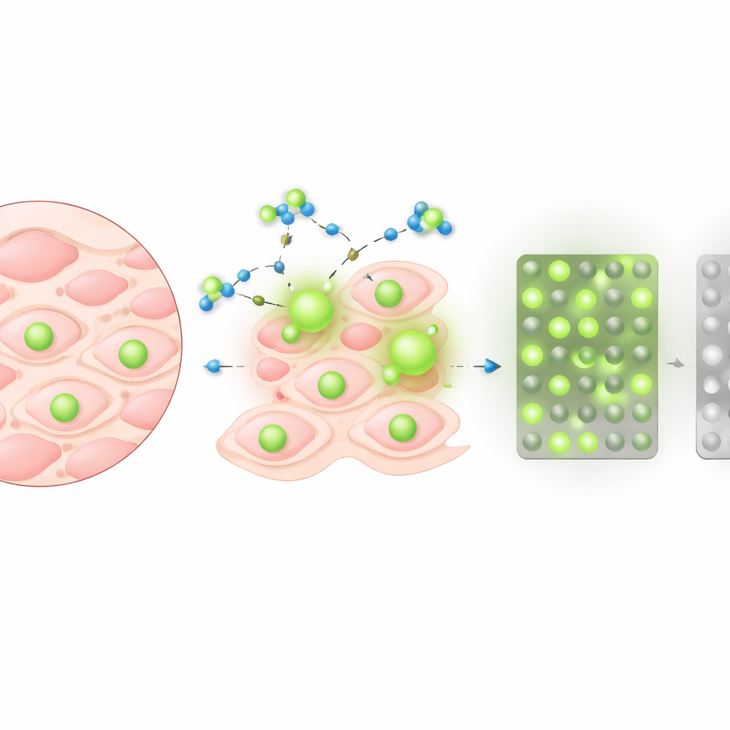
The research team developed and tested a small molecule, called the α‑Man probe, that makes breast cancer tissue fluoresce—emit light—when it interacts with a specific enzyme found at high levels in many breast cancer cells. This enzyme, α‑mannosidase 2C1, is part of how cells process sugar‑decorated proteins, and it is much more active in tumors than in normal breast tissue. By dropping the probe solution directly onto small samples cut from the removed breast tissue, the scientists could quickly record how much the tissue brightened over time. Because this method works on the same tissue that will later be examined under the microscope, it fits naturally into current surgical workflows.
Testing the idea in real surgical samples
The team first carried out an exploratory study at one breast cancer hospital. They collected 93 tissue samples from 46 patients, including invasive cancers, non‑invasive cancers, and normal or benign tissue. For each sample, they measured how much the brightness changed in the first few minutes after adding the α‑Man probe. Cancerous samples, both invasive and non‑invasive, showed a clear, time‑dependent increase in fluorescence that set them apart from normal and benign tissue. Using statistical tools, the researchers defined cut‑off values for the change in brightness after five minutes and after twenty minutes. These values allowed them to place each sample into three groups: clearly cancerous, clearly non‑cancerous, or uncertain.
Sharper answers than an earlier fluorescent test
The same group had previously created a different probe targeting another enzyme (GGT), but that earlier method often produced a confusing background glow in normal breast tissue. In head‑to‑head comparisons, the new α‑Man probe identified a much larger fraction of cancer samples as clearly positive, while keeping the rates of false alarms and missed cancers very low. In other words, fewer samples fell into the frustrating “uncertain” middle zone. At five minutes, the new probe correctly confirmed cancer in about 94% of positive readings and correctly ruled it out in about 95% of negative readings—figures that rival many established diagnostic tests.
Proving it works across multiple hospitals
To see whether this method would hold up beyond a single center, the researchers then ran a larger multicenter study involving four hospitals and 274 analyzable samples, of which 118 contained cancer. Again, cancer samples glowed more strongly than benign tissue, and the same three‑tiered system—positive, uncertain, negative—could be applied. At five minutes, the test delivered high positive and negative predictive values while leaving about half of all samples in the uncertain group. When the team re‑checked these borderline cases at twenty minutes, many shifted confidently into the clearly benign category, cutting the uncertain group down to less than 40% while slightly improving the reliability of negative results. Importantly, the probe performed similarly well across all participating hospitals and across different types of breast cancer, with the exception of mucinous tumors, which were excluded because their mucus‑rich makeup interfered with the signal.
A simple two‑step guide for surgeons
Based on these findings, the authors propose a practical flow for use during breast‑conserving surgery. After removing the tumor, surgeons or staff would apply the α‑Man probe to thin slices from the surgical margin and measure the brightness increase at five minutes. A clearly positive result would suggest that cancer cells remain at the edge and that additional tissue should be removed. A clearly negative result would support stopping the operation without further cutting, especially when the main concern is leaving behind invasive cancer. For samples that fall into the uncertain range, a second reading at twenty minutes would help separate truly benign tissue from the few remaining suspicious cases, which could then be sent for traditional frozen‑section analysis.
What this could mean for patients
For people undergoing breast‑conserving surgery, this fluorescent probe could make operations shorter, safer, and less dependent on scarce pathology resources. Although it cannot yet replace detailed microscopic examination, it offers a fast, repeatable way to check many margin samples at once, potentially lowering the need for second surgeries due to missed cancer at the edge. The authors conclude that the α‑Man probe is a promising tool for real‑time surgical guidance and plan future clinical trials to confirm whether its use can reliably reduce false‑negative margins and re‑operation rates in everyday practice.
Citation: Ueo, H., Ueo, H., Doi, T. et al. Multicenter ex vivo evaluation of an α-mannosidase–targeted fluorescent probe for intraoperative margin assessment in breast cancer. Sci Rep 16, 11789 (2026). https://doi.org/10.1038/s41598-026-42316-2
Keywords: breast cancer surgery, fluorescence imaging, surgical margin assessment, intraoperative diagnostics, molecular probes