Clear Sky Science · en
Enhancing lung cancer classification through a double attention hybrid CNN-HiFuse approach
Why Finding Lung Cancer Early Matters
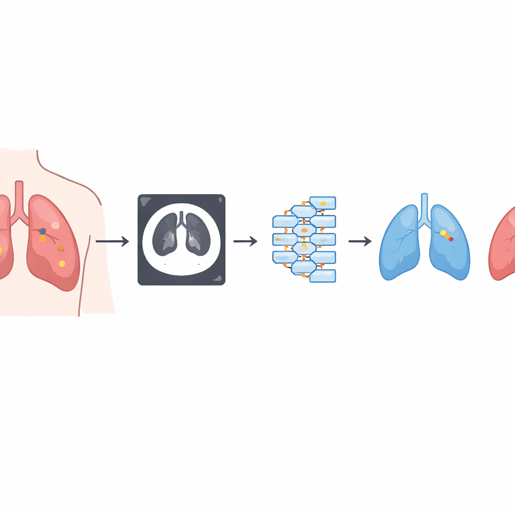
Lung cancer is one of the deadliest cancers worldwide, in large part because it is often discovered late, when treatment options are limited. Radiologists already use detailed CT scans to search for tiny spots in the lungs that might be cancer, but carefully inspecting every image slice is slow, tiring work—and small or subtle nodules can be missed. This study explores how an advanced kind of artificial intelligence can act as a tireless second pair of eyes, helping doctors spot lung problems earlier and tell harmless findings from dangerous ones.
Turning Chest Scans into Clear Categories
The researchers focus on a practical three-way decision that radiologists face every day: does a CT slice show normal lung tissue, a harmless (benign) nodule, or a cancerous (malignant) nodule? They use a publicly available dataset from an oncology center in Iraq that contains 1,190 CT slices from 110 patients, each patient labeled as normal, benign, or malignant. To avoid overestimating performance, they split data by patient instead of by image, so slices from the same person never appear in both training and testing. They also standardize image size, brightness, and format, and apply data augmentation—such as rotations, flips, and brightness changes—to help the system cope with real-world variability and to balance the less common benign and malignant cases.
How the Smart Scan Reader Sees Images
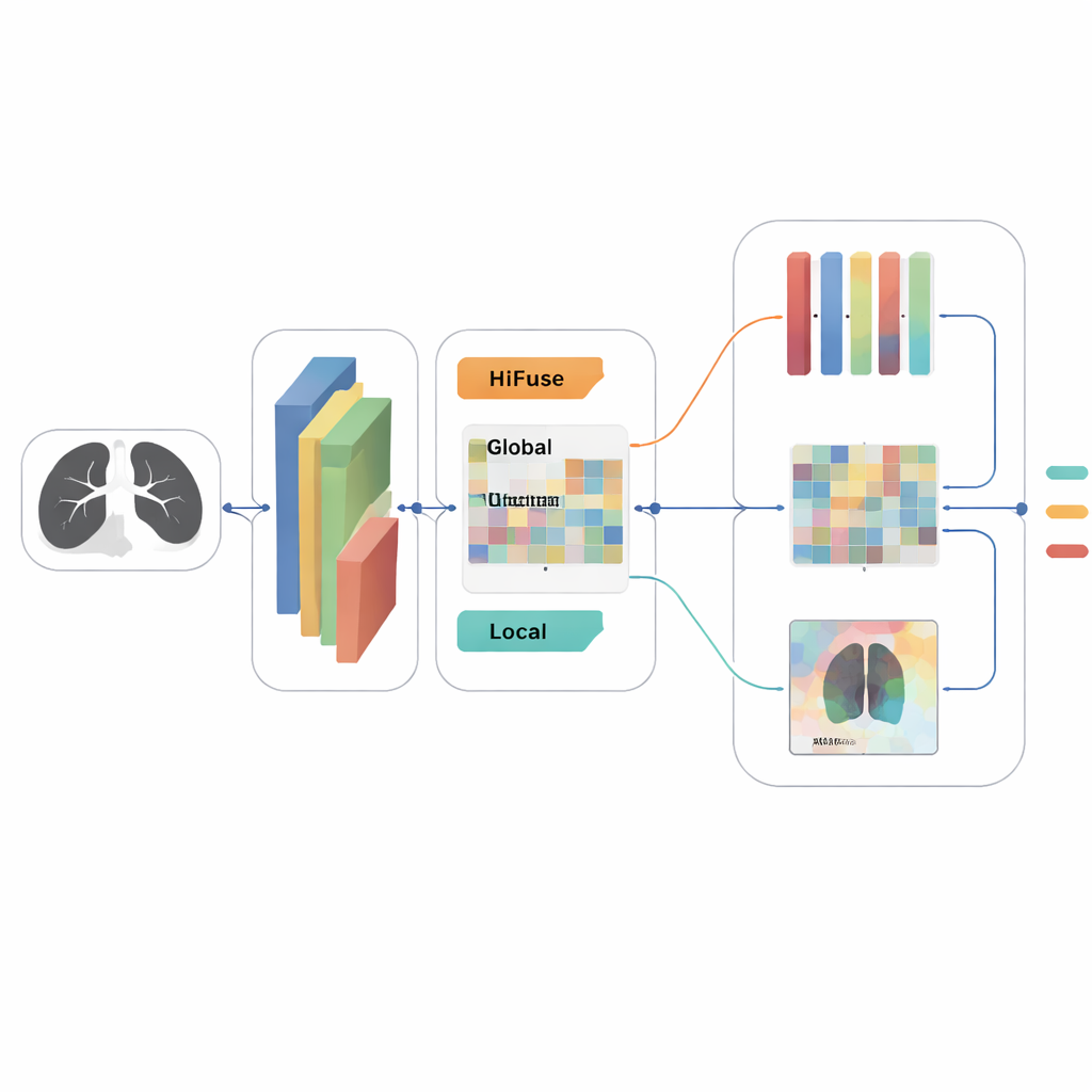
At the heart of the system is a convolutional neural network, a type of artificial intelligence especially good at recognizing patterns in pictures. The model they design, called a Double Attention Hybrid CNN–HiFuse architecture, is tailored to the challenges of lung CT. It first uses standard convolutional layers to pick up basic visual features like edges and textures. Then, a specialized module named HiFuse combines information at different scales: one branch captures broad, global context across the whole lung, while another zooms in on local details around potential nodules. By fusing these views hierarchically, the network is better able to notice both small spots and the larger structures that surround them.
Teaching the System Where to Look
Beyond simply extracting features, the model actively learns where to pay attention. A "double attention" mechanism operates in two ways. Channel attention assigns more weight to the most informative types of features—for example, those that are particularly good at distinguishing a malignant nodule from scar tissue—while downplaying distracting signals. Spatial attention, in contrast, concentrates on specific regions within the image, highlighting suspicious areas in the lung and suppressing background. These two forms of attention are applied in sequence, helping the system create a compact, focused summary of each scan slice, which is then passed to a final classifier that outputs the probabilities of normal, benign, or malignant tissue.
How Well the Approach Performs
To tune the many choices involved in building such a system—like how many filters to use, how quickly to adjust its internal weights, and how strongly to regularize the network—the authors employ an automated search tool. With the best settings in place, they compare their hybrid model against several strong baselines, including well-known deep networks like VGG16 and ResNet50, as well as a customized network with attention. On an independent test set of 213 CT slices, their Double Attention Hybrid CNN–HiFuse model achieves about 98 percent accuracy, with very high precision and recall. It misclassifies only four slices in total and shows particularly low rates of missing malignant nodules, an especially important safety concern. Receiver operating characteristic curves, which measure how well the model separates classes across different thresholds, approach ideal performance for all three categories.
Promise and Limits for Real-World Care
For non-specialists, the main message is that this study demonstrates a fast, relatively lightweight AI system that can classify lung CT images into three clinically meaningful groups with impressive accuracy, while also providing attention maps that show which regions most influenced its decision. This makes the tool more interpretable and easier for radiologists to trust as a decision-support aid rather than a black box. However, the results come from a single hospital, rely on 2D slices instead of full 3D scans, and use a simplified three-class labeling scheme, so they should be viewed as an encouraging first step rather than definitive proof. With further testing on larger, more diverse datasets and eventual integration into clinic workflows, similar attention-driven models could help catch lung cancer earlier and reduce the burden on overworked imaging specialists.
Citation: M. D, A., B, V. Enhancing lung cancer classification through a double attention hybrid CNN-HiFuse approach. Sci Rep 16, 13099 (2026). https://doi.org/10.1038/s41598-026-42290-9
Keywords: lung cancer, CT imaging, deep learning, computer-aided diagnosis, attention mechanisms