Clear Sky Science · en
The microbiome of interstitial cystitis revealed by 2bRAD-M
Why Bladder Pain and Tiny Tenants Matter
For many people, constant bladder pain and the need to urinate frequently can turn everyday life into a struggle. This condition, known as interstitial cystitis or bladder pain syndrome, is common in middle‑aged women yet remains poorly understood and hard to treat. The study behind this article asks a deceptively simple question with big implications: which microbes quietly live inside the bladder wall of these patients, and could they be linked to their pain? Using an advanced DNA‑based method, the researchers mapped this hidden community of microscopic tenants in bladder tissue itself, rather than in urine, opening a new window on a mysterious disease. 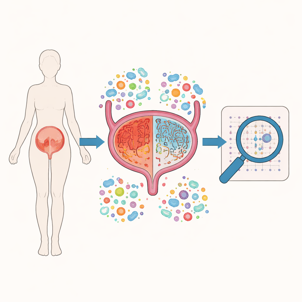
A Closer Look at a Painful Condition
Interstitial cystitis/bladder pain syndrome causes long‑lasting pain or pressure in the pelvis, together with urgency, frequent urination, and waking up at night to urinate. Doctors know that the bladder lining often shows signs of irritation and inflammation, but they do not agree on what triggers it. Earlier work hinted that microbes might play a role: some patients improve with antibiotics, and their urine or stool sometimes shows altered bacterial communities. Still, studies of urine alone have not revealed a clear “signature” germ for this condition. The authors of this paper reasoned that the real story might lie in the bladder wall itself, where microbes could reside without always appearing in urine samples.
A New Way to Read Microbial Fingerprints
To peer into this hidden world, the team used a technique called 2bRAD‑M, which reads tiny, uniquely shaped DNA fragments cut from microbial genomes. Unlike many standard tests that usually identify microbes only at the broader group level, this method can distinguish species with high precision, even when only traces of DNA are available. The researchers collected small pieces of tissue from 11 women with interstitial cystitis: one sample from visibly reddened, “lesion” areas and another from nearby regions that looked normal to the naked eye. All samples came from patients who had not recently taken antibiotics or had urinary infections, reducing the chance that obvious outside factors distorted the microbial picture.
What Lives Inside the Bladder Wall
Across 22 tissue samples, the team detected DNA from 118 bacterial species and two fungi. Overall, the microbial communities in painful lesion areas and in neighboring normal‑looking tissue were remarkably alike. Both contained a mix dominated by a few groups of bacteria, including species related to Escherichia coli, Bacillus, and Chlamydia‑like organisms. Strikingly, three species—Mycobacterium tuberculosis, a particular Ralstonia strain, and Klebsiella pneumoniae—were found in every single tissue sample. These microbes are known from other diseases, ranging from tuberculosis to urinary infections and even some cancers, but their exact role here is unclear: they could be long‑term residents, harmless bystanders, or contributors to a smoldering irritation of the bladder lining.
Small Differences and Hidden Activities
When the team compared lesion tissue to nearby normal‑appearing tissue, they found that overall diversity—the number of different species and how evenly they were represented—did not differ much. This supports the idea that, in this form of the disease, the whole bladder may be affected rather than having sharply confined trouble spots. Even so, subtle differences emerged. Certain microbes, such as those in the Sphingopyxis group and the Rhizobiaceae family, were more common in lesion tissue, while others, including Acetobacteraceae and Porphyromonas, were relatively enriched in the normal‑looking areas. Using computer tools to predict what these microbes might be doing, the researchers found that key metabolic pathways—such as those handling fatty acids, core energy production, and vitamin‑related compounds—were shifted between lesion and normal tissues, hinting that microbial activity could influence inflammation and tissue health. 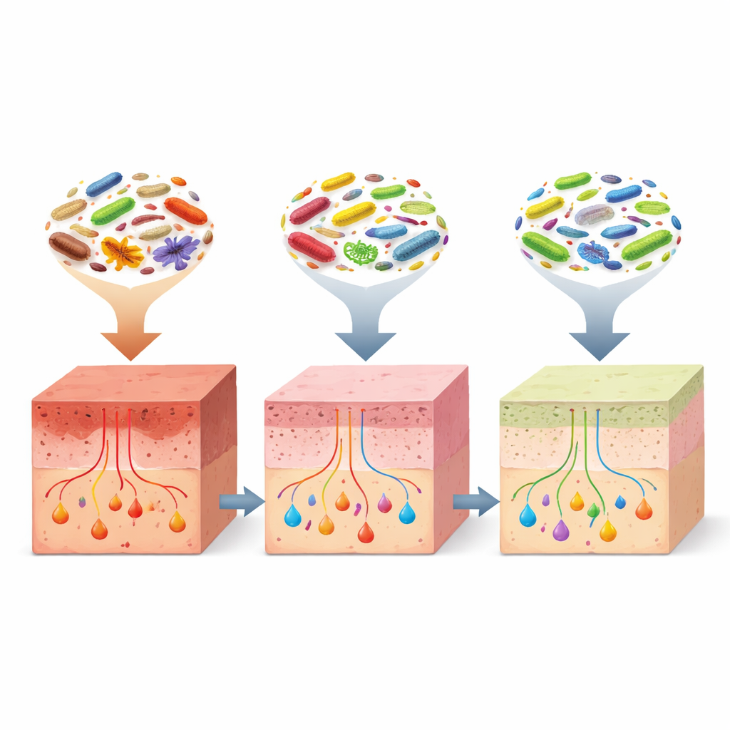
What This Means for Patients and Future Research
To a lay reader, the key message is that the bladder wall in people with interstitial cystitis is not sterile: it hosts a fairly stable community of microbes, and this community looks broadly similar in painful and nearby normal‑looking regions. This supports the idea that the disease involves widespread, diffuse changes rather than isolated patches of damage. The discovery of three common bacterial species in all samples and of a few groups that differ subtly between lesion and normal tissue offers new clues, but not yet clear villains. More research with healthy and disease controls will be needed to learn whether these microbes help cause the condition, arise as a result of it, or simply coexist with it. Still, by revealing the hidden residents of the bladder wall, this study provides a new starting point for understanding chronic bladder pain and, eventually, for designing more targeted treatments.
Citation: Gan, Y., Zhang, J., Yao, K. et al. The microbiome of interstitial cystitis revealed by 2bRAD-M. Sci Rep 16, 12413 (2026). https://doi.org/10.1038/s41598-026-42249-w
Keywords: interstitial cystitis, bladder microbiome, chronic pelvic pain, tissue-resident bacteria, microbial sequencing