Clear Sky Science · en
Quantitative analysis of myeloid cell patterns and immunosuppressive enzyme (IDO, ARG1) expression in colorectal cancer pulmonary metastases and corresponding primary tumours
Why the Body’s Own Defenses Sometimes Help Lung Tumors
When colorectal cancer spreads to the lungs, doctors want to know which patients are most likely to benefit from surgery to remove those lung spots. This study looks closely at the body’s immune cells inside these lung tumors and asks a puzzling question: why do some immune cells that normally weaken the anti-cancer response sometimes seem linked to better survival, while in other situations they predict a worse outcome?
Two Enzymes That Quiet the Immune System
The researchers focused on two immune-dampening enzymes, called IDO and ARG1. These molecules are produced by certain white blood cells and sometimes by cancer cells themselves. They work by breaking down amino acids that T cells—the front-line soldiers of the immune system—need to grow and function. Because of this, IDO and ARG1 are often viewed as helpers of the tumor, and new drugs are being tested to block them. Yet earlier work in primary colorectal tumors in the bowel had revealed a surprise: higher levels of these enzymes were often linked with better survival. The new study asks whether the same is true once cancer has spread to the lungs.
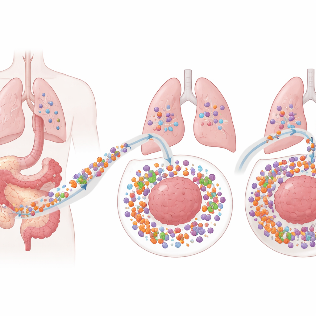
Zooming In on Cells in Lung Metastases
The team studied 91 lung metastases from 53 people with colorectal cancer, comparing them with the patients’ original bowel tumors. Using highly multiplexed staining and machine-learning image analysis, they could identify different kinds of myeloid cells (a family of immune cells that includes monocytes and granulocytes), their “maturity,” and whether they carried IDO or ARG1. They sampled both the outer edge of each tumor, where cancer meets normal lung (the invasive margin), and the deeper tumor center. This allowed them to build a detailed map of which cells, in which locations, were associated with how long patients lived after lung surgery.
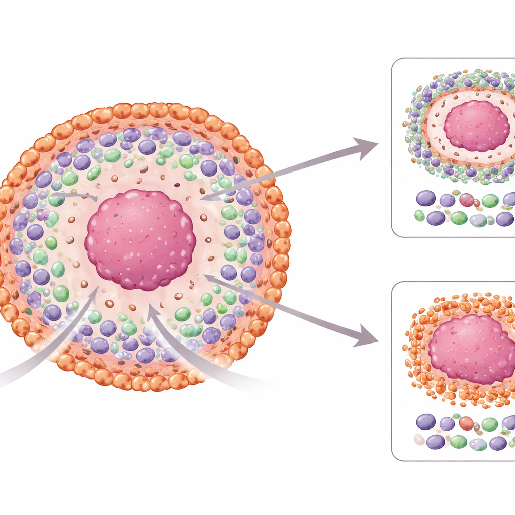
Good News in the Tumor Center, Warning Signs at the Edge
The results showed that location mattered. In the center of the lung tumors, higher numbers of IDO-carrying monocytes were linked with clearly better five-year survival, even after accounting for age, treatment, and the presence of T cells. These helpful cells tended to look more “mature” and carried another surface feature, FCGR3, suggesting they may be better at presenting tumor pieces to the immune system and keeping cancer under control. In contrast, at the invasive margin, more monocytes overall, and especially monocytes either lacking IDO or carrying IDO while still immature, were associated with shorter survival. Certain ARG1-carrying granulocytes in and around the tumor center also pointed toward worse outcomes, especially in patients who had not received chemotherapy before surgery.
Different Immune Landscapes in Lung and Bowel Tumors
When the lung metastases were compared to the original bowel tumors, important differences emerged. The edges of the lung tumors contained more myeloid cells, showed higher levels of IDO and ARG1, and had more cells expressing FCGR3, hinting at a more strongly shaped immune environment. IDO-carrying monocytes sat closer to cancer cells than their IDO-negative counterparts, while ARG1-negative granulocytes clustered nearer the tumor than ARG1-positive ones, implying distinct ways these cell types interact with cancer. Notably, the patterns that previously predicted good outcomes in primary bowel tumors did not simply carry over to the lung metastases, underscoring that the immune “rules” change as cancer spreads.
What This Means for Future Treatments
For patients and clinicians, these findings suggest that not all IDO or ARG1 activity is equal. In the center of lung metastases, IDO-bearing monocytes may reflect a vigorous, organized immune response that helps patients live longer. At the growing edge of the tumor, however, immature IDO-positive monocytes and certain ARG1-positive granulocytes seem to signal a strongly suppressive environment that favors cancer progression. Understanding these spatially distinct patterns could help refine which patients might benefit from drugs targeting these enzymes and guide the design of combination therapies that account for where, and in which cells, immune suppression is happening.
Citation: Karjula, T., Elomaa, H., Väyrynen, S.A. et al. Quantitative analysis of myeloid cell patterns and immunosuppressive enzyme (IDO, ARG1) expression in colorectal cancer pulmonary metastases and corresponding primary tumours. Sci Rep 16, 11770 (2026). https://doi.org/10.1038/s41598-026-42097-8
Keywords: colorectal cancer metastasis, tumor immune microenvironment, myeloid cells, indoleamine 2,3-dioxygenase, arginase-1