Clear Sky Science · en
Müller glia derived EVs promote neurite recovery of an enriched population of retinal ganglion like cells derived from hESC retinal organoids after damage
Why eye support cells matter for vision loss
Diseases like glaucoma slowly kill the nerve cells that carry visual information from the eye to the brain, leading to permanent vision loss. Current treatments mostly aim to lower eye pressure, but they do little to directly protect or repair these fragile nerve cells. This study explores a surprising helper: support cells in the retina called Müller glia, and the tiny particles they release, as a possible way to shield and even partly repair damaged eye nerves.
Tiny packages with a big job
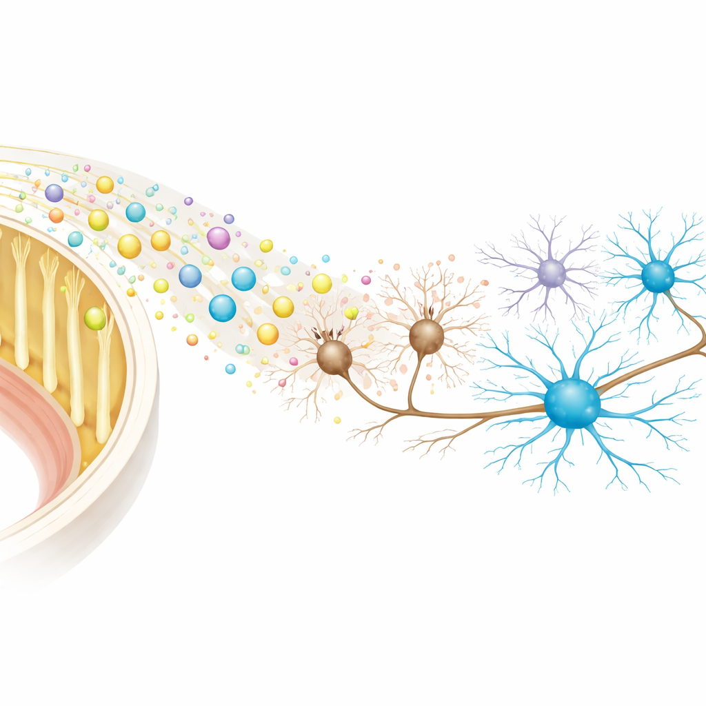
Müller glia are long, column-like cells that run through the thickness of the retina and help keep all other retinal cells healthy. The researchers focused on the microscopic bubbles these cells release, called extracellular vesicles. These vesicles are like biological parcels filled with proteins, fats and short RNA molecules that can alter how nearby cells behave. Because they are naturally produced, stable and less likely to trigger immune reactions, such vesicles are being actively explored as next-generation treatments for disorders of the brain and eye.
Building a human lab model of eye nerve cells
To test whether Müller-cell vesicles can protect vision-related nerve cells, the team first needed a human cell model. They grew three-dimensional “retinal organoids” from human embryonic stem cells—miniature, simplified retinas in a dish. From organoids that were 40 to 50 days old, they isolated clusters of neurons enriched for retinal ganglion cells, the type that dies in glaucoma. These clusters showed the telltale shapes and markers of retinal ganglion cells, although they also contained some other retinal neuron types, making them a realistic but not perfectly pure sample.
Hurting the cells, then trying to help them
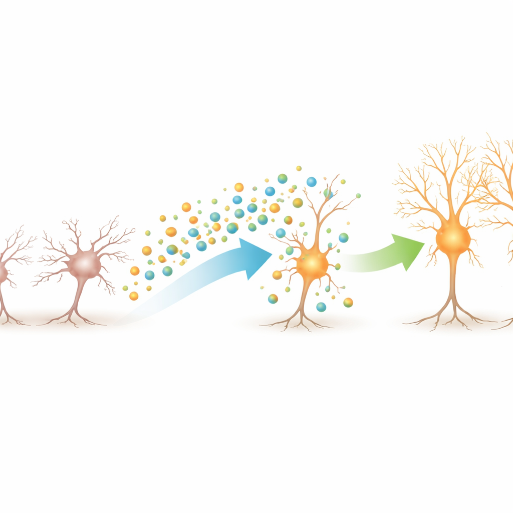
The scientists then mimicked injury by exposing these retinal ganglion–like cultures to a chemical called NMDA, which overstimulates nerve cells and shortens their long, cable-like projections known as neurites. After 24 hours of this damage, some cultures received Müller-cell vesicles, while others received only a simple salt solution, a known mix of protective growth factors, or a drug that blocks NMDA’s harmful action. By measuring neurite length from many cell clusters, they found that NMDA clearly stunted the cells’ projections. Adding Müller-cell vesicles after this damage substantially restored neurite length, to a degree similar to the established growth factor cocktail. The number of neurites per cluster did not change much, indicating that the main effect was on regrowth and extension rather than on forming new branches.
Signals that tilt the balance toward survival
To understand how these vesicles might be working inside the cells, the team examined a wide panel of signaling proteins whose activity switches on and off through chemical tags. NMDA injury alone boosted several stress and death-associated pathways, including key members of the MAP kinase family and the protein p53. When vesicles were added after the damage, this stress signature was dampened, while other pathways linked to cell survival and neurite growth, particularly a group of proteins called RSK kinases and related growth regulators, became more active. Imaging experiments confirmed that the vesicles were indeed taken up by the nerve cells, clustering around their bodies and extensions, suggesting a direct delivery of helpful cargo.
What this could mean for future eye treatments
Put simply, this study shows that tiny, naturally produced bubbles from retinal support cells can help damaged human-like eye nerve cells regrow their connections in the lab, and that they nudge the cells’ internal signals away from self-destruction and toward repair. While much work remains—such as pinpointing which vesicle components are most important, proving long-term safety, and testing in living eyes—the findings support the idea that Müller-cell vesicles could one day be harnessed as a targeted, cell-free therapy to slow or prevent vision loss in conditions like glaucoma.
Citation: Eastlake, K., Lamb, W.D.B., Tracey-White, D. et al. Müller glia derived EVs promote neurite recovery of an enriched population of retinal ganglion like cells derived from hESC retinal organoids after damage. Sci Rep 16, 11853 (2026). https://doi.org/10.1038/s41598-026-42089-8
Keywords: glaucoma, retinal ganglion cells, Müller glia, extracellular vesicles, retinal organoids