Clear Sky Science · en
WTAM-YOLO: a YOLOv11-based method for pulmonary nodule detection
Why Finding Tiny Spots in the Lungs Matters
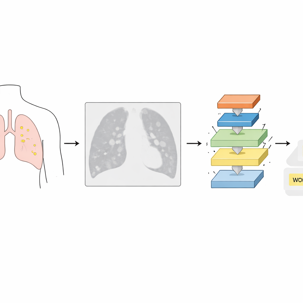
Lung cancer often begins as tiny spots called nodules that appear in medical scans long before symptoms arise. Catching these nodules early can mean the difference between a poor outlook and a high chance of long-term survival. But these specks are small, faint, and easy to confuse with normal lung structures, even for experienced radiologists and conventional computer programs. This study introduces WTAM-YOLO, a new artificial intelligence system designed to spot these elusive nodules in lung CT images more accurately and efficiently than current tools.
The Challenge of Seeing What Barely Stands Out
In a chest CT image, lung tissue appears mostly in shades of gray, with blood vessels, airways, and other structures overlapping in complex ways. Cancerous nodules can be only a few millimeters wide, sometimes little more than a soft blur. Traditional detection methods, whether human or machine, struggle to separate these subtle spots from their surroundings. Existing AI systems, including earlier versions of the popular YOLO family of object detectors, often miss very small nodules or mistake normal anatomy for disease, producing both missed cancers and false alarms.
A Smarter Way to Read CT Images
WTAM-YOLO builds on a recent YOLO design but reshapes how the computer "looks" at CT images. Instead of relying solely on standard image filters, the authors weave in several specialized components. One uses wavelets—mathematical tools that can zoom in on sharp edges and fine textures while still seeing the bigger picture. Another set of modules acts like a series of spotlight operators, automatically emphasizing regions and details most likely to contain nodules and dimming irrelevant background. Together, these parts help the system keep track of both tiny specks and the broader lung context in a single pass.
How the New System Learns Tiny Details
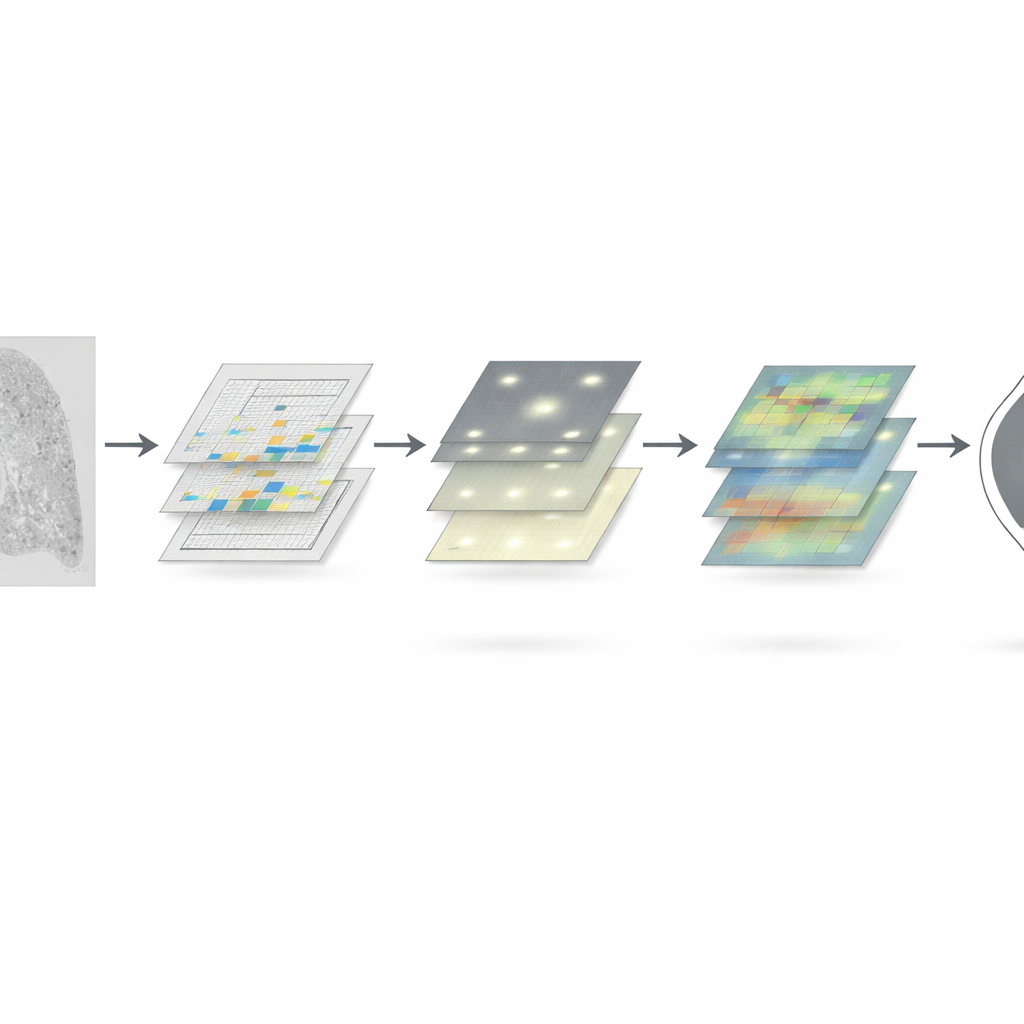
Inside WTAM-YOLO, the wavelet-based blocks first split the image information into multiple scales, capturing broad outlines and delicate edges at the same time before stitching them back together. Attention modules then reweight this information along two axes: across different types of features and across locations in the image, so that suspicious spots stand out more strongly. Additional components are designed specifically to protect the fragile signal of very small nodules as the image is repeatedly shrunk and processed. One such block smooths and stabilizes the features over training time, helping the model avoid overreacting to random noise while still remaining sensitive to tiny lesions.
Putting WTAM-YOLO to the Test
The researchers trained and evaluated WTAM-YOLO on two well-known collections of lung CT scans: a curated public dataset from the Roboflow platform and the widely used LUNA16 dataset. They compared its performance against the underlying YOLOv11 model and several other modern detectors. Across both datasets, the new system detected a higher fraction of true nodules while also reducing the number of false findings. On key measures that summarize detection quality, WTAM-YOLO delivered several percentage points of improvement, especially at stricter matching conditions that reflect precise localization—exactly the kind of rigor needed in early cancer screening.
What This Means for Patients and Clinics
To a non-specialist, the core message is that WTAM-YOLO helps computers "see" tiny lung nodules more clearly in CT scans, making them brighter against a noisy background without overwhelming doctors with false alarms. By better preserving small, subtle details and organizing information across multiple scales, the system improves both sensitivity and accuracy while keeping computing demands modest enough for practical use. Although it still relies on slices rather than full 3D scans and will need further testing in real-world hospital settings, this work points toward faster, more reliable lung nodule screening that can support radiologists and potentially catch lung cancer earlier, when treatment is most effective.
Citation: Lan, Y., Zhang, Y., Xu, J. et al. WTAM-YOLO: a YOLOv11-based method for pulmonary nodule detection. Sci Rep 16, 10922 (2026). https://doi.org/10.1038/s41598-026-42029-6
Keywords: lung nodule detection, CT imaging, deep learning, YOLO, early lung cancer