Clear Sky Science · en
Comparative evaluation and validation of rapid quantification methods for Mycoplasma hyopneumoniae: development of a PMA-based viability qPCR assay
Why Faster Germ Tests Matter for Pigs and People
Hidden in a pig’s lungs, a tiny germ called Mycoplasma hyopneumoniae quietly causes chronic cough, slower growth, and major financial losses for farmers around the world. Yet counting how many of these germs are truly alive in a sample can take weeks with older lab methods, delaying decisions about treatment, vaccination, and herd management. This study shows how a suite of modern tools, capped by a new rapid DNA-based test, can shrink that waiting period to just a few hours while focusing specifically on live, disease‑causing bacteria.
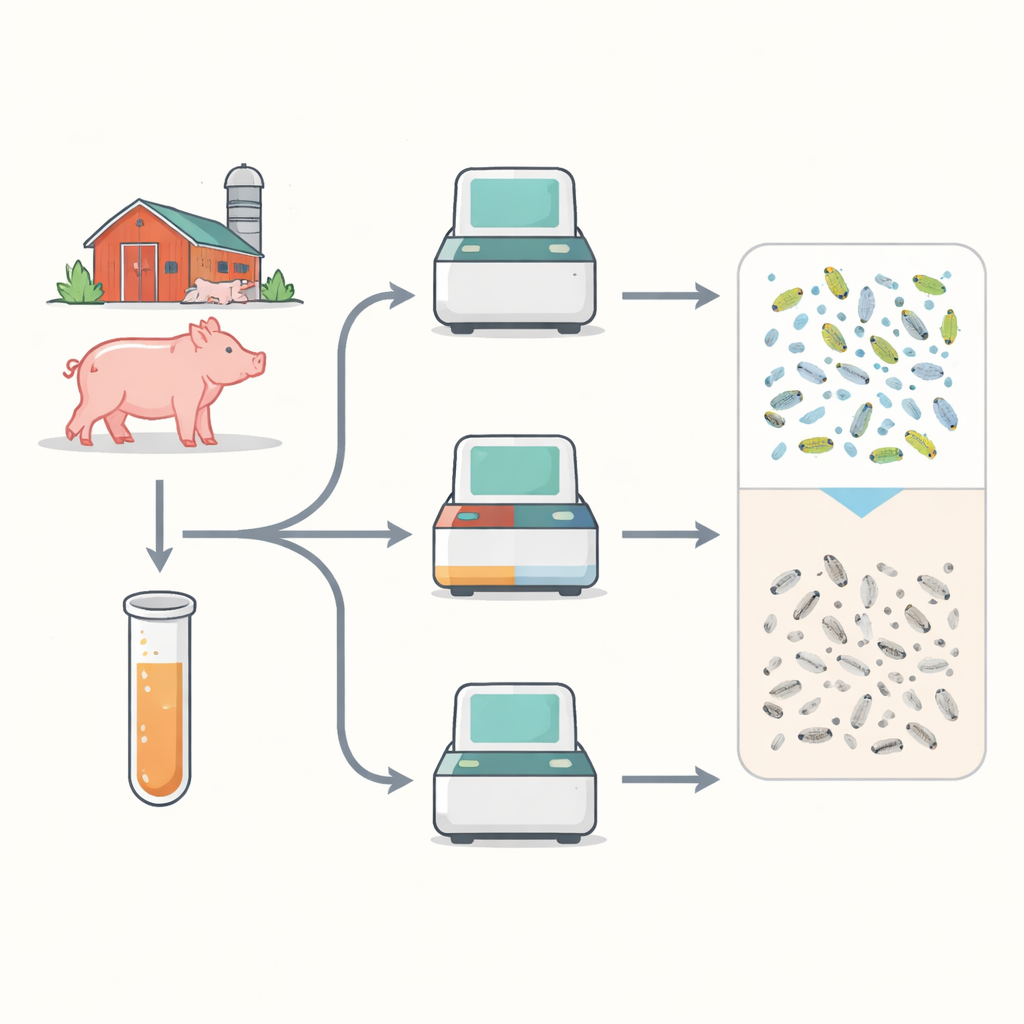
Old Ways of Counting That Take Too Long
For decades, laboratories have measured this pig lung germ using culture methods that rely on color changes in the growth liquid or the appearance of tiny colonies on agar plates. These approaches, while familiar, are slow and often unreliable: it can take two to four weeks before enough growth appears to judge how many bacteria were present, and some field strains grow so poorly on plates that they are badly underestimated. The researchers began by systematically comparing these traditional techniques with two faster laboratory readouts that track bacterial metabolism and cell integrity, asking whether the quicker tests truly tell the same story about how the germ grows.
Modern Tools That See Cells in New Ways
The team grew three representative strains of the germ and followed their progress in the lab using four approaches in parallel: the classic color-change method, colony counts on plates, a light‑based readout of cellular energy molecules, and flow cytometry, which quickly counts single cells and distinguishes live from dead ones based on fluorescent stains. They also built a confocal microscopy method that reconstructs three‑dimensional views of stained bacteria in droplets of culture liquid, serving as a second way to gauge live and dead cells. Across strains, the newer methods showed growth curves that rose and fell at similar times to the old ones, and statistical comparisons revealed mostly moderate to high agreement. Flow cytometry and confocal imaging, in particular, aligned well for two of the three strains, supporting flow cytometry as a solid reference for live cell numbers even when colonies are hard to grow.
Turning a DNA Test into a Live‑Germ Detector
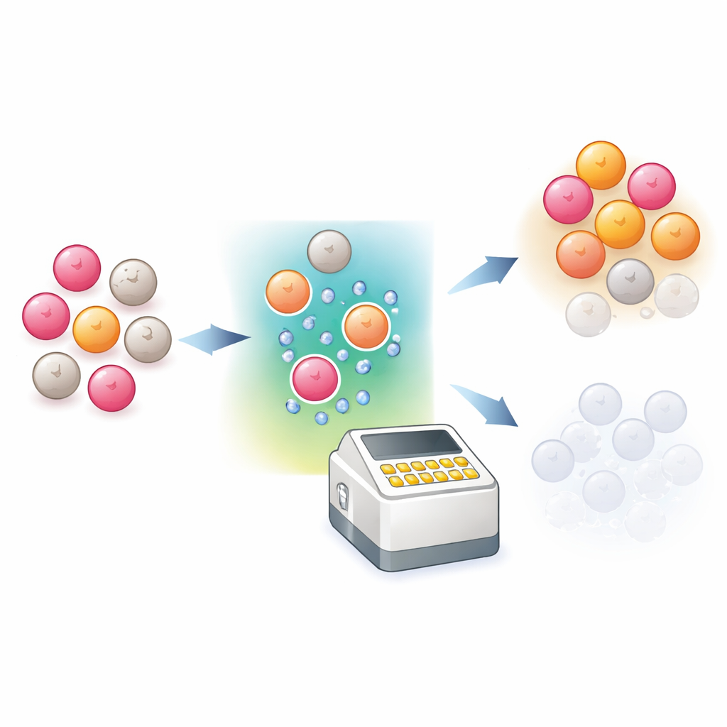
Most veterinary labs already rely on a DNA test called qPCR to detect this germ, but standard qPCR cannot tell whether the DNA comes from living cells or from dead remnants lingering after treatment or disinfection. To bridge that gap, the scientists adapted a “viability qPCR” strategy that adds a dye called propidium monoazide (PMA) before the DNA test. This dye can slip only into damaged, dead cells, where—after a burst of light—it locks onto their DNA and prevents it from being copied during qPCR. The group systematically fine‑tuned washing steps, sonication to break up clumps, shaking speed, light exposure time, and dye concentration to maximize blocking of dead‑cell signals while leaving live cells unaffected. They then tested the optimized recipe on mixes of live and heat‑killed bacteria, using flow cytometry as an independent yardstick of how many cells were truly alive.
How Well the New Test Tracks Real Viability
With the new PMA‑based protocol in place, the viability qPCR results closely mirrored the live cell counts measured by flow cytometry across all three strains, showing that the test is sensitive to changes in the number of viable bacteria. In contrast, regular qPCR produced nearly the same readout even when the proportion of live cells changed dramatically, confirming that it mainly reflects total DNA. The team also determined how few live cells the new assay can reliably detect: for the best‑characterized strain, it could still distinguish changes down to about fifty thousand live bacteria per milliliter, below which the response leveled off. Although this limit is higher than that of standard qPCR, it reflects the extra challenge of silencing the DNA signal from large numbers of dead cells in the background.
What This Means for Pig Health and Farm Decisions
By combining fast cell‑counting methods with a carefully tuned viability‑focused DNA test, this work offers veterinarians and producers a practical way to know not just whether the germ is present, but whether it is truly alive in meaningful numbers—and to learn that in hours instead of weeks. Once the new assay is fully validated on real‑world samples such as swabs and lung washes from pigs, it could sharpen herd‑level decisions about when to treat, how well control programs are working, and whether animals used to introduce immunity into a herd are carrying mostly live or mostly dead bacteria. In the broader sense, this study shows how pairing smart chemistry with existing DNA tests can transform them from simple presence‑or‑absence tools into more informative gauges of active infection.
Citation: Ko, C.C., Gauger, P.C., Rigby, S. et al. Comparative evaluation and validation of rapid quantification methods for Mycoplasma hyopneumoniae: development of a PMA-based viability qPCR assay. Sci Rep 16, 11504 (2026). https://doi.org/10.1038/s41598-026-41951-z
Keywords: Mycoplasma hyopneumoniae, swine respiratory disease, viability qPCR, flow cytometry, diagnostic methods