Clear Sky Science · en
Structure and diversity of abdominal exocrine glands in larvae of Leiodidae (Insecta: Coleoptera: Staphylinoidea)
Tiny Larvae with Hidden Superpowers
On the surface, the pale, soft-bodied larvae of small scavenger beetles look like unremarkable grubs quietly feeding on decaying animals. But under the microscope, their skin hides a sophisticated system of tiny glands that may help them survive in some of the filthiest, microbe-filled places on Earth. This study peels back the layers of that hidden machinery, revealing how these larvae are equipped with intricate abdominal glands whose secretions may protect them, help them stick to surfaces, or play other roles in their lives.
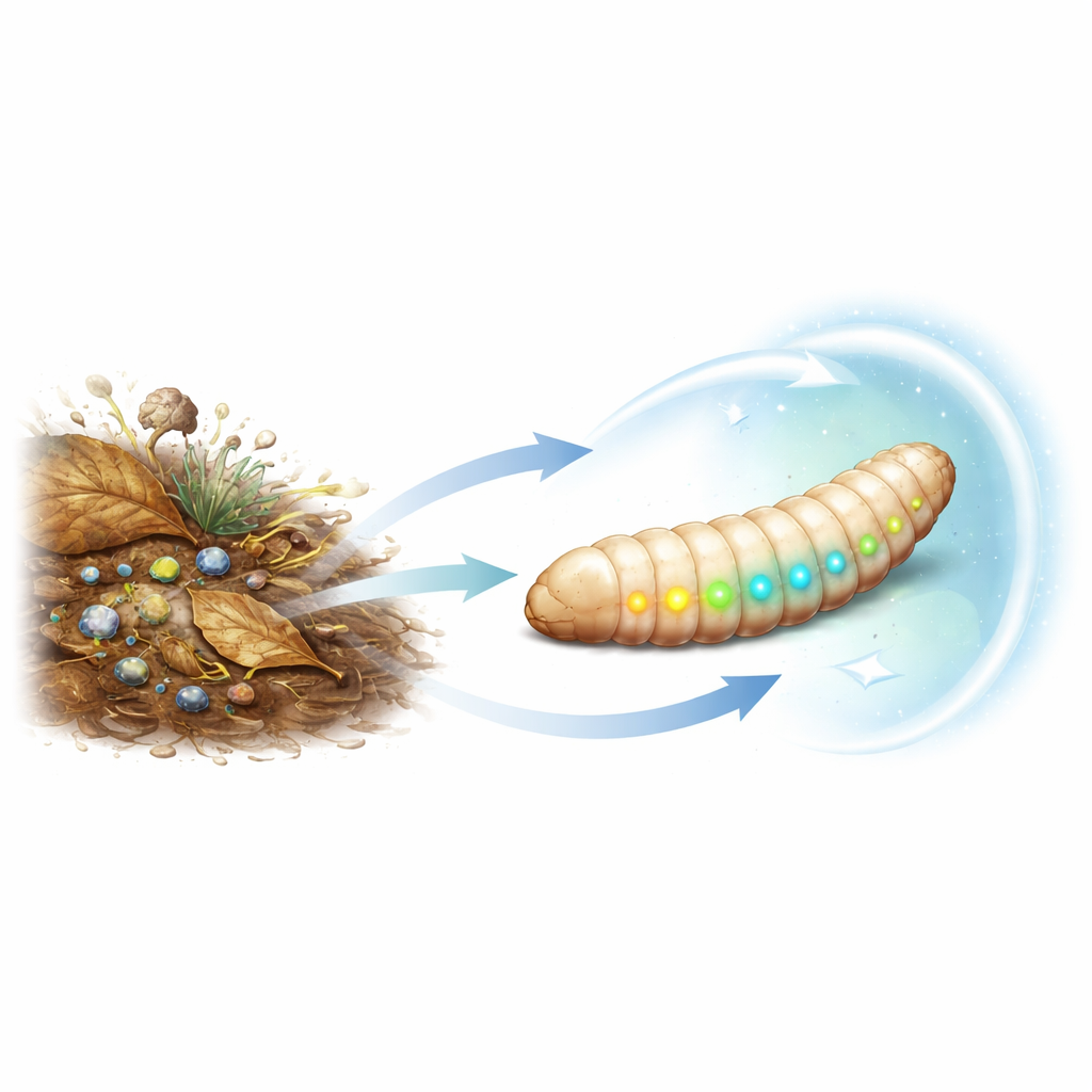
Life in Rotting Worlds
The beetles at the center of this work belong to the families Leiodidae and Agyrtidae, groups of scavengers that live in damp habitats rich in decaying animal matter, fungi, and bacteria. Such environments are both a feast and a health hazard: food is abundant, but so are pathogens and predators. Adult beetles from many families are known to possess exocrine glands—specialized structures that release chemicals to the outside of the body. These secretions can repel enemies, communicate with other beetles, or keep the body surface clean. Much less is known, however, about similar glands in the larval stages, even though larvae spend most of their time buried in these risky habitats.
A New Look at Larval Skin
The researchers focused on larvae of the scavenger beetle Sciodrepoides watsoni watsoni, examining all three growth stages using a suite of powerful imaging tools. Light microscopes showed that each abdominal segment carries a pair of small, complex structures on the upper side. Scanning electron microscopy revealed that each of these complexes combines several kinds of openings in the outer shell: a raised oval area peppered with hundreds of fine pores, a central flower-like (rosette) opening, and several cup-like openings nearby. Transmission electron microscopy then allowed the team to look inside, where they found three distinct types of glandular cells arranged between the outer shell and the body muscles.
Three Glands Working Side by Side
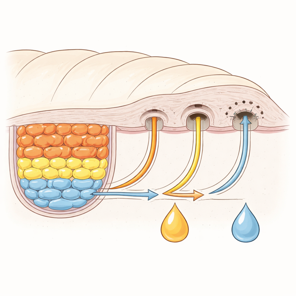
Inside each segmental complex, one group of long, narrow cells sends finger-like projections up to the base of the perforated oval plate. Their tips are packed with tiny surface folds, suggesting they produce material that is released through the roughly 300 microscopic pores. A second set of larger cells forms a kind of internal pouch: their upper surface folds inward to create a reservoir-like space lined with dense surface projections. Here, secretion gathers and is funneled outward through a shallow “cup” and then through a cup-shaped opening in the outer shell. A final, very large cell works together with a canal-forming partner cell to build a tiny duct that ends at the rosette-like opening. This unit is crammed with energy-producing structures and secretion-filled pockets, hinting at an active production of complex substances. Notably, each gland type has its own dedicated outlet; their secretions do not merge before reaching the surface.
Variety Across the Beetle Family Tree
To understand how widespread and diverse these glands are, the authors compared larvae from many species of Leiodidae and Agyrtidae, drawing on both new material and past descriptions. They found that similar abdominal gland complexes occur in most of the species examined, but with striking differences in layout and shape. In some larvae, the perforated plate lies in front of a small ridge on the segment; in others it lies behind it, or the ridge is missing altogether. The perforated area can be oval, bean-shaped, or elongated, and the number and type of openings range from a single pair to as many as twenty. These patterns line up in intriguing ways with current ideas about how different subgroups of these beetles are related, suggesting that the glands may carry useful clues for reconstructing their evolutionary history.
What Are These Secretions For?
Although the study does not yet identify the chemistry of the secretions, their cellular machinery and the larvae’s lifestyle point to likely roles. Species such as S. watsoni live on decaying animal remains where harmful microbes thrive, and the larvae are coated in a stubborn film that is hard to wash away. From other beetle groups, similar glands are known to produce antimicrobial coatings, sticky defensive fluids, or materials used for building shelters and cocoons. The combination of storage spaces, ducts, and energy-rich cells seen here fits well with the idea of an external protective or adhesive layer tailored to life in wet, dirty habitats.
Why These Hidden Glands Matter
By mapping these tiny structures in detail for the first time in this beetle family, the study shows that larvae are far from simple “wormlike” life stages. Instead, they carry elaborate glandular equipment that likely helps them defend themselves and manage contact with their challenging surroundings. The diversity of gland designs across related species also offers a new set of traits that can help biologists untangle beetle family relationships and trace how different lifestyles evolved. In short, what looks like a plain grub to the naked eye turns out, under the electron microscope, to be a finely engineered organism equipped with an entire chemical toolkit built into its skin.
Citation: Kilian, A., Garbiec, A., Růžička, J. et al. Structure and diversity of abdominal exocrine glands in larvae of Leiodidae (Insecta: Coleoptera: Staphylinoidea). Sci Rep 16, 12426 (2026). https://doi.org/10.1038/s41598-026-41930-4
Keywords: beetle larvae, exocrine glands, insect defence, ultrastructure, scavenger beetles