Clear Sky Science · en
Risk factors for post-traumatic osteoarthritis following surgical treatment of acetabular posterior wall fractures: a retrospective study
Why Hip Injuries Matter Long After the Crash
When someone survives a serious car crash or a fall from a height, the immediate concern is often broken bones and emergency surgery. But for many, the real trouble begins years later, when a damaged hip joint slowly wears out and becomes painfully stiff — a condition known as post‑traumatic osteoarthritis. This study looks at a specific kind of hip socket break, called a posterior wall acetabular fracture, and asks a practical question: after surgeons repair the break, which early warning signs tell us who is most likely to develop crippling arthritis and eventually need a hip replacement?
The Broken Edge of the Hip Socket
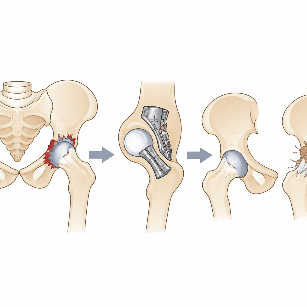
The hip joint works like a ball in a cup: the ball is the top of the thigh bone, and the cup is the bony socket of the pelvis. In high‑energy accidents, the back edge of that cup can shatter, often combined with the ball popping out of place. Surgeons can realign the bone and hold it with plates and screws, but this does not guarantee a healthy joint in the long run. Earlier research on hip socket fractures in general hinted at links between severe breaks, damaged joint surfaces, and later arthritis, yet there was little focused evidence for this particular back‑wall break. The authors of this study set out to fill that gap, hoping to pinpoint which details of the original injury and the surgery matter most for long‑term hip health.
Following Patients from Operation to Outcome
The team reviewed records from two major trauma centers in China, including 159 adults who had only the back wall of the hip socket broken and were treated with open surgery to reset and fix the bone. All operations used a standard approach from the back of the hip, and patients were followed for at least two years, with regular scans and a widely used hip function questionnaire. To count as post‑traumatic osteoarthritis, patients had to have both symptoms (such as pain and poor hip score) and clear X‑ray signs of joint wear, including narrowed space between the ball and socket and bony overgrowths. The researchers then compared dozens of factors — from age, bone quality, and smoking to the exact fracture pattern, timing of surgery, and quality of the repair — to see which ones were tied most strongly to later arthritis.
What Turned a Fixed Hip into an Arthritic Hip
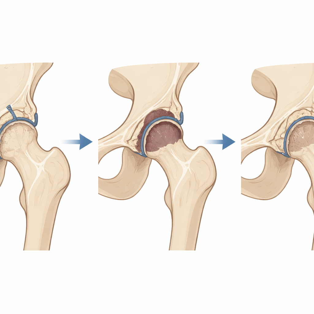
Nearly one in four patients (23.9%) went on to develop post‑traumatic osteoarthritis, usually within about a year and a half, and roughly a third of those ultimately needed a total hip replacement. After sorting through the data, several patterns stood out. Hips with more shattered bone pieces (comminution), or in which the joint surface had been crushed inward like a dented plate, were more likely to deteriorate. If a large portion of the back wall — at least half of the socket’s arc — was missing, the risk climbed further, likely because the ball was no longer well contained. Perhaps most striking, patients whose thigh‑bone head later lost its blood supply and began to die (femoral head necrosis) had the largest jump in risk, reflecting how crucial a living, stable ball is to a lasting joint.
Timing and Precision in the Operating Room
The way surgery was done, and when, also played a major role. When surgeons achieved a near‑perfect match between the broken pieces — restoring the smooth curve of the cup — patients were less likely to develop arthritis. In contrast, even small residual steps or gaps hastened wear by disturbing how forces spread across the cartilage. Operations that ran longer were also linked to higher arthritis risk, probably because they represented more complicated, severely damaged hips that required extensive handling of bone and soft tissues. Another clear signal was timing: patients operated on more than two weeks after injury had a markedly higher likelihood of later arthritis than those treated earlier, suggesting that delays allow cartilage damage and subtle deformities to become harder to reverse.
What This Means for Patients and Surgeons
To a patient, the message of this study is simple but powerful: with this type of hip socket fracture, the seriousness of the initial damage and the details of the repair strongly shape the joint’s future. Severe shattering, a heavily crushed joint surface, a large missing back wall, imperfect realignment, loss of blood supply to the hip ball, long operations, and delayed surgery all push the joint toward early arthritis and possible hip replacement. Conversely, getting to surgery sooner and restoring the cup’s smooth shape as precisely as possible improve the odds of keeping one’s natural hip. These findings give doctors clearer checklists for judging risk, planning surgery, and counseling patients about both the urgency of treatment and the long‑term outlook for their injured hip.
Citation: Yuan, G., Ke, X., Lian, J. et al. Risk factors for post-traumatic osteoarthritis following surgical treatment of acetabular posterior wall fractures: a retrospective study. Sci Rep 16, 11210 (2026). https://doi.org/10.1038/s41598-026-41893-6
Keywords: hip fracture, post-traumatic osteoarthritis, acetabular fracture, hip replacement, orthopedic surgery