Clear Sky Science · en
Validation of AI-enhanced ECG image analysis for identifying extreme cardiac magnetic resonance metrics in a cross-ethnic UK biobank study
Why a simple heart test could do much more
Most people know the electrocardiogram, or ECG, as the quick test where stickers and wires record the heart’s electrical signals. What if those familiar squiggles could reveal early signs of serious heart trouble long before symptoms appear—without expensive scanners or long hospital visits? This study explores how artificial intelligence (AI) can turn ordinary ECG recordings into a powerful screening tool that spots people with hidden heart damage usually seen only on advanced MRI scans.
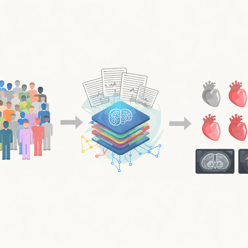
Turning everyday heart traces into deeper insight
Traditional ECGs are cheap, fast, and available almost everywhere, but they have limits: many structural heart problems do not leave obvious clues in the standard readout. Advanced imaging tests, such as cardiac magnetic resonance (CMR) scans, give far more precise pictures of heart size, shape, and pumping strength—but they are costly, time‑consuming, and not widely available. The researchers behind this paper previously built AI systems that can “look” at ECG images and estimate subtle heart problems that doctors normally confirm with ultrasound or MRI. Here, they set out to test whether those AI tools still work well in a large, general population very different from the hospital patients on which they were first trained.
A massive health database as a real‑world test bed
The team used data from the UK Biobank, a long‑running project that has collected health information, including scans, from hundreds of thousands of volunteers. They focused on 38,804 people who had both a standard 12‑lead ECG and a CMR scan taken at the same visit. Instead of looking only for clear‑cut disease, the researchers defined “abnormal” hearts as those with the most extreme 1% of MRI measurements—hearts that pumped poorly, showed signs of strain, had unusually thick muscle, or had an enlarged upper chamber. This allowed them to ask a simple question: can AI that reads ECG images reliably pick out the small fraction of people whose MRI scans show worrisome changes?
How well the AI spotted weak or enlarged hearts
The six AI‑ECG models performed impressively. They were especially good at detecting weakness of the main pumping chamber on the left side of the heart, reaching accuracy levels that earlier work had only shown in smaller or more uniform groups of patients. The systems also did well at recognizing subtle problems on the right side of the heart and in identifying people whose heart muscle was unusually thick or whose left upper chamber was enlarged. Across the board, people flagged by AI tended to be older and more likely to have high blood pressure or other risk factors, matching what doctors would expect. The AI’s performance stayed strong across different age groups, body sizes, and medical histories, and in many situations it outperformed traditional ECG rules that doctors have used for decades.
What this could mean for everyday care
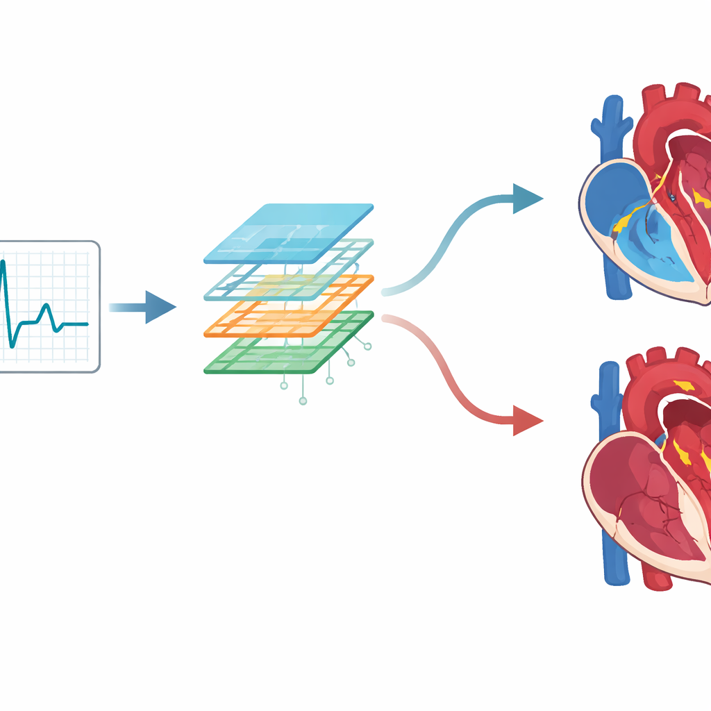
Because the AI works on standard ECG images, it could be added to existing machines or software with minimal extra equipment. In a clinic, pharmacy, or community screening event, an ECG could quietly run through the AI model in the background, highlighting a small number of people whose hearts look especially abnormal and who might benefit from a closer look with MRI or ultrasound. The models were particularly good at ruling out major problems: if the AI score was low, it was very unlikely that the person had one of the extreme MRI findings. That makes this approach well‑suited as an early warning filter to focus scarce imaging resources where they are most needed.
Important caveats and the road ahead
The study does have limits. Most volunteers in the UK Biobank are of European background and generally healthier than typical hospital patients, so the results may not fully reflect performance in more diverse or sicker populations. The definition of “abnormal” was based on statistical extremes rather than standard clinical cutoffs, which may not match how doctors usually diagnose disease. And like many modern AI systems, these models function largely as black boxes, offering little direct explanation for how they reach a decision. The authors stress that future work must test the tools prospectively, follow patients over time, and improve transparency before AI‑ECG can be woven safely into everyday practice.
A simple test with far‑reaching promise
Overall, the study shows that AI can extract rich information about heart structure and function from an ordinary ECG, closely matching what is seen on sophisticated MRI scans in a large, community‑based group. For a layperson, the message is straightforward: a routine heart tracing, when paired with smart algorithms, could one day serve as an early radar system for hidden heart problems. If carefully validated and deployed, AI‑enhanced ECGs might help doctors catch trouble sooner, guide who needs more advanced tests, and extend high‑quality heart screening to places where complex imaging is out of reach.
Citation: Kim, Y., Lee, H., Choi, HM. et al. Validation of AI-enhanced ECG image analysis for identifying extreme cardiac magnetic resonance metrics in a cross-ethnic UK biobank study. Sci Rep 16, 11996 (2026). https://doi.org/10.1038/s41598-026-41824-5
Keywords: AI electrocardiogram, cardiac MRI, heart failure screening, left ventricular hypertrophy, population heart health