Clear Sky Science · en
Subclinical myocardial dysfunction in treatment naive papillary thyroid carcinoma patients
Hidden Heart Risks from a Common Thyroid Cancer
Papillary thyroid carcinoma is one of the most curable cancers, and many patients live long, healthy lives after diagnosis. Yet this study asks a quietly unsettling question: even before any surgery or hormone treatment, could the cancer itself be nudging the heart toward trouble in ways that standard tests miss? Using advanced heart imaging and blood markers, the researchers looked for very early, "silent" changes in heart function in people newly diagnosed with papillary thyroid cancer.
Why Look Beyond Survival?
Because papillary thyroid cancer usually has an excellent outlook, attention is increasingly shifting from whether patients survive to how well they live over decades. Large population studies have hinted that people with well-differentiated thyroid cancers may face higher rates of heart rhythm problems, heart failure, and coronary artery disease than the general public, even after accounting for usual risk factors. Until now, most of that concern has focused on the side effects of long-term thyroid-stimulating hormone (TSH) suppression therapy. The present study asks whether the disease itself, before any treatment, might already be reshaping the heart in subtle ways that routine scans fail to capture. 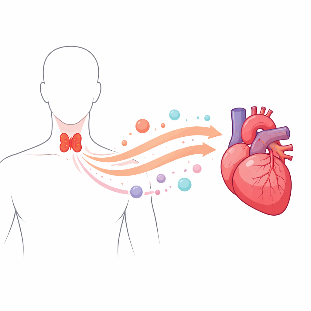
Who Was Studied and How
The researchers enrolled 36 adults with newly diagnosed papillary thyroid cancer who had not yet received surgery, radiation, or thyroid hormone pills, and compared them with 20 patients who had benign nodular goiter. All participants had normal thyroid blood tests and similar age, sex, body size, blood pressure, cholesterol levels, and neck artery thickness, which helped isolate the impact of cancer itself. The team used a battery of sophisticated ultrasound techniques to examine the heart. In addition to standard measures such as ejection fraction—the proportion of blood the heart pumps with each beat—they used tissue Doppler imaging and speckle-tracking echocardiography to measure how the heart muscle relaxes and how its fibers shorten and stretch, captured in a metric called global longitudinal strain.
Early Heart Changes That Routine Tests Miss
On the surface, the hearts of cancer and non-cancer patients looked similar: ejection fraction remained in the normal range for both groups, and basic size measurements did not differ meaningfully. But the more sensitive tests told a different story. More than half of the papillary thyroid cancer patients showed mild diastolic dysfunction—meaning the heart muscle relaxed more sluggishly between beats—compared with only a quarter of those with benign nodules. Their global longitudinal strain was also measurably worse, indicating early weakness in how the heart muscle fibers contract, despite overall pumping strength still appearing normal. These findings suggest that by the time papillary thyroid cancer is first diagnosed, the heart may already be experiencing subtle functional strain.
Clues from Blood Markers and Hormone Sensitivity
The team then turned to the bloodstream for clues about what might be linking thyroid cancer to these early heart effects. They measured a cell-surface receptor called integrin αvβ3, which is known to be abundant on thyroid cancer cells and on cells involved in new blood vessel growth and heart tissue repair. Levels of this molecule were clearly higher in cancer patients than in those with benign nodules and were even higher in patients whose cancer had spread to lymph nodes. Importantly, higher integrin αvβ3 levels tracked with worse global longitudinal strain and with a greater likelihood of mild diastolic dysfunction, even after accounting for age, sex, and body mass index. The researchers also assessed a measure of how sensitively the brain’s control centers respond to thyroid hormone signals, called the Thyroid Feedback Quantile-based Index. This index was higher—indicating reduced central sensitivity—in the cancer group and was linked both to higher integrin αvβ3 levels and to poorer heart strain measurements. Together, these patterns hint at a web of interactions among thyroid cancer biology, hormone signaling, and the heart. 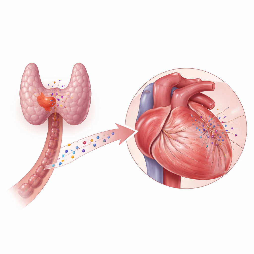
What This Means for Patients
To a non-specialist, the key message is that even in patients who feel well, have normal thyroid blood tests, and show a normal ejection fraction, papillary thyroid cancer may already be leaving faint fingerprints on the heart. These fingerprints appear as small but detectable changes in how the heart relaxes and how its fibers contract—changes that are tied to both a cancer-related receptor in the blood and to altered sensitivity of the brain–thyroid feedback loop. The study does not prove that these early shifts will inevitably lead to heart disease, nor does it show that treating the cancer will worsen or improve them. However, it raises the possibility that simple blood markers and advanced ultrasound techniques could one day help flag thyroid cancer patients who are quietly more vulnerable to cardiovascular problems and who might benefit from closer long-term heart monitoring.
Citation: Akin, S., Akgul, G.G., Gulcelik, M.A. et al. Subclinical myocardial dysfunction in treatment naive papillary thyroid carcinoma patients. Sci Rep 16, 12439 (2026). https://doi.org/10.1038/s41598-026-41816-5
Keywords: papillary thyroid cancer, subclinical heart dysfunction, echocardiography, integrin avb3, thyroid hormone sensitivity