Clear Sky Science · en
Astrocyte diversity and aging in the mouse lemur primate brain
Why tiny primates matter for brain aging
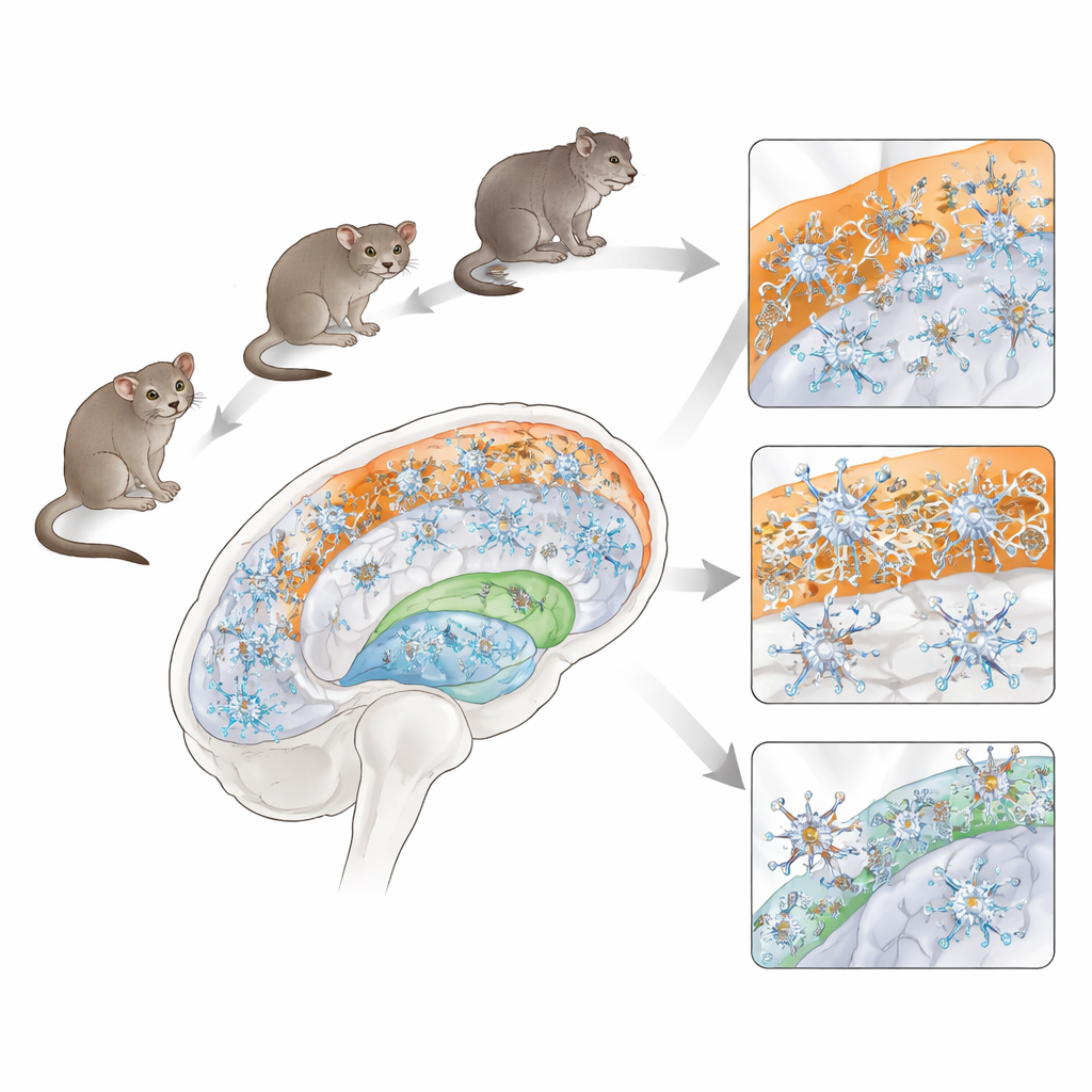
As we age, our brains do not just lose neurons; the support cells that keep those neurons healthy also change. Among the most important of these helpers are astrocytes, star-shaped cells that regulate blood flow, clear chemicals, and maintain the brain’s delicate balance of salts and water. Most of what we know about astrocytes comes from rodents, yet human astrocytes are more varied and complex. This study turns to an unusual model—the gray mouse lemur, a tiny primate that ages quickly—to explore how different types of astrocytes are organized across the brain and how they change with age in a species closer to us than mice.
Mapping the brain’s hidden caretakers
The researchers examined the brains of 17 gray mouse lemurs ranging from young adults to the equivalent of human centenarians. Using tissue staining methods that highlight astrocytes, they created a whole-brain map of where these cells live and what they look like. Astrocytes were especially abundant in the brain’s internal wiring, known as white matter, and in the hippocampus, a region important for memory. In contrast, the main thinking surface of the brain, the cortex, contained surprisingly few astrocytes in its depths; most cortical astrocytes clustered near the boundary with white matter or along the outer surface of the brain.
Many shapes, many roles
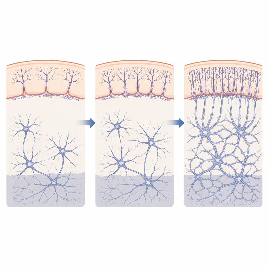
Within this map, the team identified a rich variety of astrocyte shapes that likely reflect different jobs. In white matter, “fibrous” astrocytes formed networks aligned with bundles of nerve fibers and were often wrapped around blood vessels, suggesting roles in maintaining nerve insulation and blood–brain exchange. In the hippocampus, “protoplasmic” astrocytes formed dense, sponge-like territories that touched many synapses while still keeping clear boundaries from one another. At the very outer surface of the cortex, they found striking vertical arrays of “interlaminar” astrocytes whose long, straight processes descend from the brain’s surface down through several layers, forming palisade-like columns seen mainly in primates and some carnivores. They also observed specialized radial cells in the hypothalamus, including tanycytes along the walls of the third ventricle, extending long processes deep into the tissue.
Beaded branches as a warning sign
A recurring feature across several astrocyte types was the presence of bead-like swellings, called varicosities, decorating their long processes. These appeared in projection astrocytes that span cortical and hippocampal layers, in interlaminar astrocytes at the surface, in tanycytes lining the brain’s fluid spaces, and in border cells at the base of the hypothalamus. Two patterns emerged: continuous chains of beads along a branch, and interrupted, more fragmented beads. Earlier work in humans has linked such beaded astrocytes to aging and disease. The widespread, sometimes fragmented varicosities seen here suggest that many astrocyte types may pass through altered physiological states as the brain ages, potentially reflecting stress or adaptation rather than a single uniform response.
Where aging hits hardest
By comparing middle-aged and old lemurs, the authors found that astrocyte aging is highly uneven across the brain. White matter showed the most dramatic changes: in older animals, astrocytes there were more numerous, larger, and more densely packed, with thicker branches, indicating strong “reactivity” often associated with tissue stress. The density and size of these cells rose together, pointing to robust structural remodeling. In contrast, deeper cortical layers and the hippocampus showed only modest overall changes. One notable exception was the interlaminar astrocytes at the cortical surface, whose descending processes became denser in older lemurs, suggesting that this primate-specific astrocyte type is particularly sensitive to aging. There was also marked variation between individuals—some very old lemurs had relatively quiet astrocytes, while others showed pronounced reactivity.
What this means for human brain health
To a layperson, the main message is that brain aging is not a simple, uniform decline. In this small primate, the support cells that help nerve fibers and surface layers function are especially vulnerable, while other regions stay relatively stable unless disease strikes. The diversity of astrocyte types in the gray mouse lemur, and their similarity to those in larger primates, make this species a powerful bridge between rodent experiments and human brain aging. By showing that white matter and surface astrocytes are key hotspots of age-related change, the work points researchers toward where to look for early signs of decline—and where future therapies might best support the aging brain.
Citation: Garcia, L., Dupuis, L., Petit, F. et al. Astrocyte diversity and aging in the mouse lemur primate brain. Sci Rep 16, 13482 (2026). https://doi.org/10.1038/s41598-026-41759-x
Keywords: astrocytes, brain aging, white matter, mouse lemur, glial cells