Clear Sky Science · en
Phase-targeting rapid cryofixation of the beating heart and histological analysis unveil contractile state-dependent sarcomere dynamics
Freezing the Heart in Mid-Beat
The human heart beats about 100,000 times a day, yet we have never truly seen what its microscopic machinery looks like at the exact moments of squeezing and relaxing. This study introduces a way to “freeze time” inside a beating heart, capturing the tiny contractile units of heart muscle in action. Understanding these changes could help explain how hearts pump efficiently in health and fail in conditions like arrhythmia or heart failure.
A New Way to Stop Motion Without Stopping Life
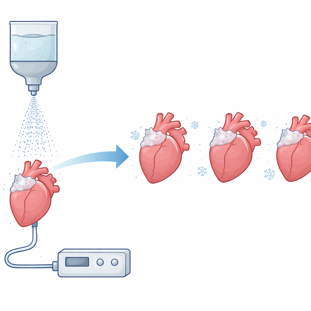
To look inside a working heart, scientists must stop its motion without giving its cells time to change shape. Traditional chemical fixatives, such as formaldehyde, spread slowly through tissue, blurring the difference between contraction and relaxation. The authors built a system that perfuses an isolated rat heart so it continues beating outside the body and then blasts its surface with an ultra-cold liquid, rapidly freezing it at a chosen moment in the heartbeat. By precisely timing this cryogenic spray to electrical pacing of the heart, they could capture tissue at peak squeeze (systole), full relaxation (diastole), or even during chaotic beating known as ventricular fibrillation.
Looking at the Heart’s Tiny Contractile Units
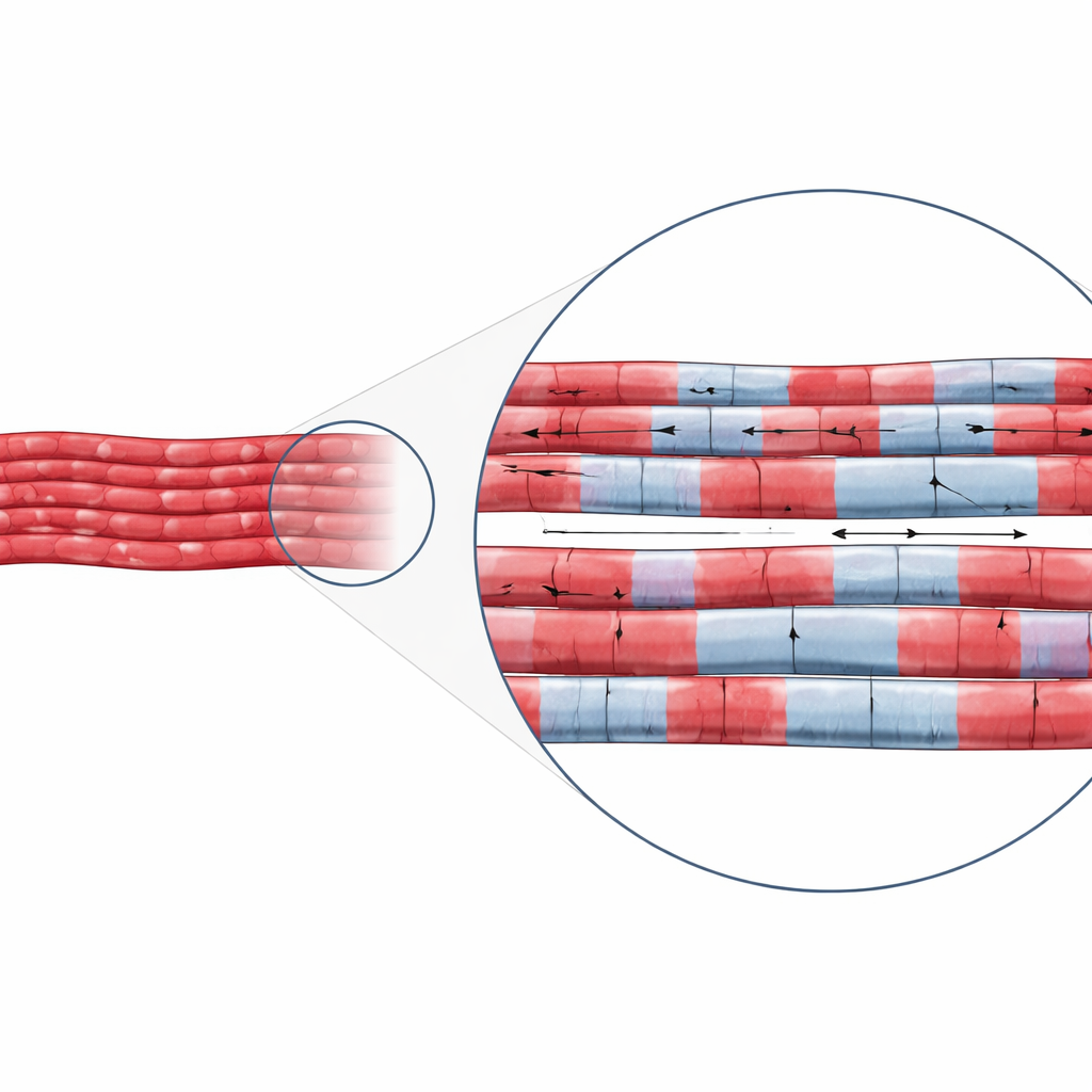
Once frozen, the hearts were gradually warmed and stabilized to preserve their microscopic structure. The researchers then used fluorescent labels to highlight key parts of the sarcomere, the repeating unit that shortens and lengthens as heart muscle contracts. They stained structures that mark the ends of each sarcomere, as well as the thin and thick filaments that slide past one another. Confocal microscopes provided detailed images from just below the frozen surface of the left ventricle, allowing the team to map how long each sarcomere was in many neighboring muscle cells at once.
Shortening, Stretching, and Patchy Behavior
The measurements confirmed a simple but fundamental rule: during systole the sarcomeres were clearly shorter than during diastole. On average, sarcomeres measured about 1.57 micrometers long when the heart was squeezing and about 1.93 micrometers when it was relaxed, consistent with earlier single-cell and small-area studies. But the frozen snapshots revealed a more complex picture than a heart that is uniformly contracted or relaxed. Even at peak squeeze, some regions contained sarcomeres that were less shortened than their neighbors. During diastole, when the heart should be relaxed, patches of still-short sarcomeres appeared among longer, more stretched ones. When the team applied a drug (BDM) that chemically relaxes the muscle, this patchiness was greatly reduced, suggesting that the uneven lengths reflected real mechanical behavior rather than freezing artifacts.
Chaos in a Quivering Heart
The approach was especially revealing during ventricular fibrillation, a dangerous rhythm in which the heart quivers instead of pumping. Live calcium imaging showed that signals inside cells became disordered, with waves of calcium rising and falling at different times across the tissue. When the researchers rapidly froze hearts in this state, the resulting sarcomere maps showed a striking mosaic of short and long segments, both within single cells and between neighboring cells. By contrast, hearts fixed more slowly with standard chemicals during fibrillation looked almost uniformly relaxed, masking the underlying chaos. This demonstrates that conventional fixation can erase critical information about how the heart fails during arrhythmias.
Why Freezing Heartbeats Matters
By stopping the heart in mid-motion with millisecond precision, this study reveals that the microscopic engine of the heartbeat is far from uniform. Sarcomeres shorten and lengthen unevenly across the heart wall, especially during relaxation and in arrhythmic states. The new cryofixation method opens a window onto these hidden patterns, providing high-resolution “snapshots” that complement live imaging. In the long run, such insights may help researchers understand why some regions of the heart become mechanically weak or unstable and guide better treatments for conditions ranging from diastolic dysfunction to life-threatening ventricular fibrillation.
Citation: Tamura, S., Mochizuki, K., Kumamoto, Y. et al. Phase-targeting rapid cryofixation of the beating heart and histological analysis unveil contractile state-dependent sarcomere dynamics. Sci Rep 16, 11484 (2026). https://doi.org/10.1038/s41598-026-41756-0
Keywords: heart muscle, sarcomere dynamics, cryofixation, ventricular fibrillation, cardiac imaging