Clear Sky Science · en
Contribution of tissue clearing and 3D image analysis to in vitro modeling of human cortical development
Why tiny model brains matter
Scientists are increasingly growing miniature, brain-like tissues in the lab to study how the human brain develops and to test new treatments for neurological diseases. But to trust what these models tell us, researchers need accurate ways to see which kinds of nerve cells they contain and how those cells are arranged in three dimensions. This study shows that moving from traditional thin tissue slices to fully three-dimensional imaging can dramatically improve how faithfully we read these tiny model brains, especially for rare or clustered cell types that 2D methods frequently miss.
From flat slices to see-through spheres
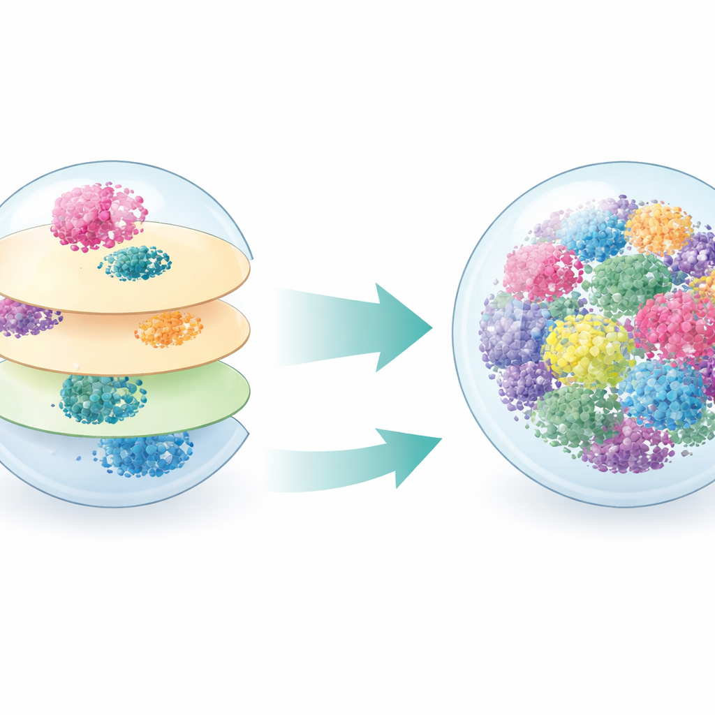
Most past work on brain organoids and neurospheres has relied on cutting samples into very thin slices and staining them under a microscope. While this two-dimensional approach is familiar and useful, it looks at only a fraction of the tissue at a time and can easily overlook cells that form small patches. The authors instead used a technique called tissue clearing, which makes whole neurospheres optically transparent while keeping their internal structure intact. Combined with light-sheet microscopy, this allowed them to image entire three-dimensional volumes and count stained cells throughout the whole sphere, rather than estimating from a few slices.
Building and imaging mini cortices
The team started with human skin cells, reprogrammed them into induced pluripotent stem cells, and then guided them to form neurospheres resembling early human cerebral cortex. Over several weeks, these spheres grew and matured, producing a mix of dividing cells and neurons resembling different layers of the cortex. The researchers stained the neurospheres with a set of well-known markers that identify specific neuron subtypes and cell states. They analyzed some samples using classic 2D cryosections and others using the iDISCO+ clearing protocol followed by 3D light-sheet imaging and computerized spot detection to count labeled cells across the full volume.
What 3D reveals that 2D misses
When the authors compared the two methods at a mid-maturation stage, they found that 2D and 3D agreed quite well for markers that are spread fairly evenly through the neurosphere, such as Ki67 (dividing cells) and CTIP2 (one class of deep-layer neurons). However, the picture changed dramatically for markers that label neurons in small clusters. For SATB2 and especially FOXP2, which highlight specific upper and deep cortical neuron subtypes, 2D slices consistently underestimated how many cells were present—by nearly an order of magnitude in the case of FOXP2. Because thin sections sample only a few planes, they often cut through the edges of clusters or miss them entirely, while 3D imaging captures every cell in context.
Following neuron growth over time
Taking advantage of the more reliable 3D approach, the researchers next tracked how different cortical neuron populations emerged as neurospheres matured from day 25 to day 60. They observed large increases in total cell number and in the absolute counts of neurons marked by BRN2 (upper layers), CTIP2 (layer V), and FOXP2 (layer VI). The spheres expanded and filled with more neurons of each type, reflecting ongoing growth and maturation. Yet when the team expressed each marker as a fraction of all cells, the proportions stayed surprisingly stable over time. This suggests that, within the time window tested, neurospheres mainly scale up while preserving a relatively constant balance of cortical neuron subtypes.
Seeing cells that share identities
The study also tested whether 3D imaging improves the measurement of cells that carry two different identity markers at once—a more demanding task. The authors focused on neurons that express both CTIP2 and COUP-TF1, a small but important group linked to specific projection patterns in the developing cortex. In thin sections, overlapping signals could be seen, but counting them across the whole neurosphere required guesswork. With 3D spot-based analysis, the team could precisely determine which labeled spots truly occupied the same space in three dimensions. This revealed nearly three times as many double-positive cells as 2D methods had suggested, underscoring how strongly partial sampling can distort our view of rare, spatially clustered populations.
What this means for brain models and therapies
Overall, the work shows that while traditional 2D histology is adequate for abundant, evenly distributed cell types, three-dimensional cleared imaging is essential for accurately capturing complex, patchy, or rare cell populations in brain-like cultures. For scientists using organoids and neurospheres to study brain development, disease, or cell therapies, this means that relying only on slices can misrepresent which cells are truly present and in what numbers. By preserving spatial integrity and measuring whole volumes, tissue clearing and 3D analysis offer a more faithful picture of how these mini brains are built, helping researchers better judge when they are ready for experimentation or transplantation and improving the reliability of findings drawn from them.
Citation: Retho, A., Govindan, A.D., Bonnet, ML. et al. Contribution of tissue clearing and 3D image analysis to in vitro modeling of human cortical development. Sci Rep 16, 13326 (2026). https://doi.org/10.1038/s41598-026-41741-7
Keywords: brain organoids, neurospheres, 3D imaging, cortical development, tissue clearing