Clear Sky Science · en
Paired regional complementarity in diffusion MRI reveals disease-specific microstructural profiles in PD, MSA, and PSP: a feasibility study
Why this brain scan research matters
Many people have heard of Parkinson’s disease, a movement disorder that can cause tremor, stiffness, and balance problems. Less well known are two related conditions—multiple system atrophy and progressive supranuclear palsy—that can look very similar in the clinic but require different care and have different outlooks. This study shows how a refined way of reading brain scans can tease these conditions apart by focusing on just a few key regions deep inside the brain, potentially helping doctors reach clearer diagnoses sooner.
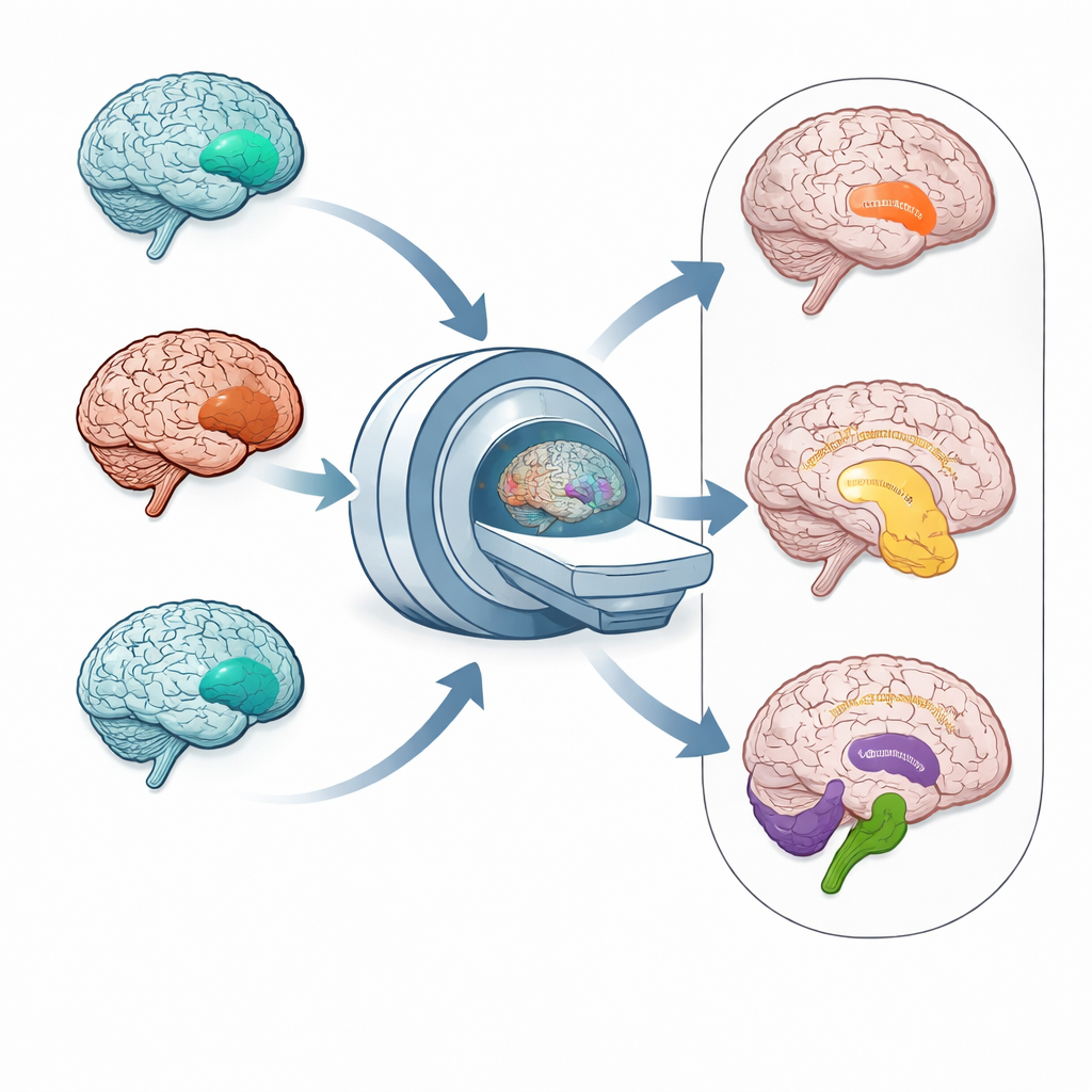
Three look‑alike diseases
Parkinson’s disease (PD), multiple system atrophy (MSA), and progressive supranuclear palsy (PSP) share overlapping symptoms such as slowed movement, stiffness, and problems with balance and thinking. Under the surface, however, they are driven by different disease processes and damage different parts of the brain. Today, even experienced specialists can struggle to tell them apart, especially early on, and standard MRI scans often show only subtle changes. A more precise and practical imaging method that highlights the right structures could make it easier to choose the most suitable treatments and to design focused clinical trials.
Listening to water movement in the brain
The researchers turned to diffusion MRI, a technique that tracks how water moves through brain tissue. Water slips more freely through damaged or thinned tissue and flows in more organized paths along healthy bundles of nerve fibers. From these patterns, scientists derive simple numerical measures, such as how strongly water prefers one direction over others, which reflects the integrity of nerve pathways. Instead of feeding dozens of these measurements from many regions into a complex, opaque computer model, the team deliberately searched for the two most informative diffusion measures in a small set of important brain areas, aiming for a compact and easily understood “signature” for each disease.
Spotlighting the brain’s traffic hubs
The study analyzed diffusion MRI data from nearly 200 patients treated for Parkinsonian syndromes. After carefully matching groups by age and sex, the team examined twelve selected brain regions, including the putamen (involved in movement control), the corpus callosum (the main bridge between the two brain hemispheres), and the cerebellum and its connecting fibers, which help coordinate movement and balance. They also studied the shape of these structures and traced major nerve pathways to see where brain tissue had thinned or nerve fibers had dwindled.
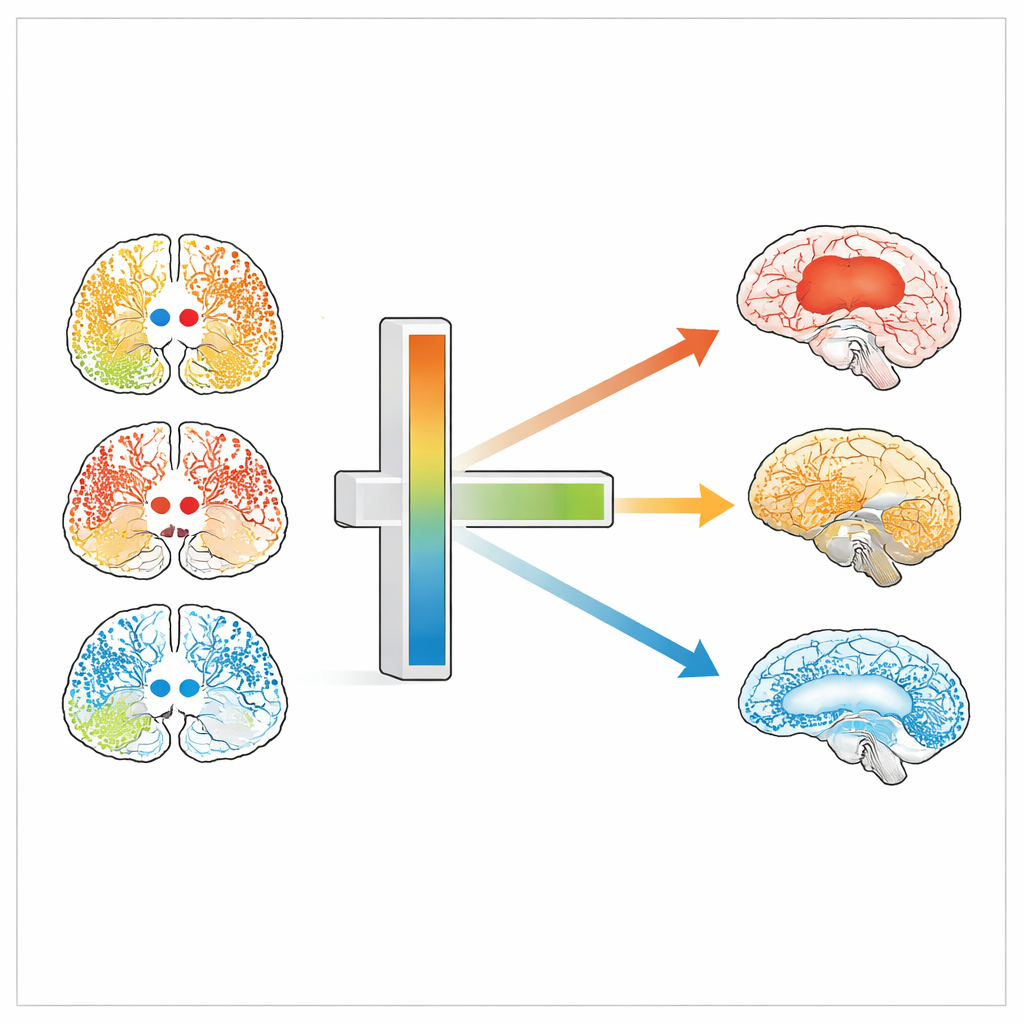
Distinct patterns for each condition
Clear, disease‑specific patterns emerged. In MSA—especially the subtype dominated by balance and coordination problems—the cerebellum and nearby pons showed marked shrinkage, looser water motion, and weaker directional flow, all signs of severe tissue damage. In PSP, the main damage involved the superior cerebellar peduncle and the corpus callosum, key highways linking movement and thinking regions. In contrast, PD showed its strongest changes in the putamen and nearby structures, with altered diffusion and fewer nerve fibers there but relatively milder damage elsewhere. By pairing just two diffusion measures from two regions—for example, how organized the fibers are in the cerebellar white matter alongside those in the putamen—the researchers could separate PD, PSP, and MSA with surprisingly high accuracy, without resorting to complex black‑box algorithms.
From complex scans to clearer decisions
To a non‑specialist, the core message is that different Parkinson‑like diseases leave different “footprints” in the brain’s wiring, and those footprints can be detected by watching how water moves through a few key hubs. This work shows that a small, carefully chosen set of diffusion MRI measurements can distinguish PD, PSP, and MSA in a transparent way that doctors can understand and trust. While larger, multi‑center studies are still needed before this approach becomes routine in hospitals, it points toward future brain scans that do not just show a blurry picture, but also offer clear, biologically grounded clues about which specific disease a patient has and how best to manage it.
Citation: Tessema, A.W., Jo, S., Kim, Y.R. et al. Paired regional complementarity in diffusion MRI reveals disease-specific microstructural profiles in PD, MSA, and PSP: a feasibility study. Sci Rep 16, 11841 (2026). https://doi.org/10.1038/s41598-026-41695-w
Keywords: Parkinsonian syndromes, diffusion MRI, brain microstructure, multiple system atrophy, progressive supranuclear palsy