Clear Sky Science · en
A novel risk-scoring system based on endoscopic ultrasound and clinical characteristics for the preoperative diagnosis of small gastric gastrointestinal stromal tumors
Why tiny stomach lumps matter
Many people discover small lumps under the lining of the stomach during routine endoscopy, often done for heartburn or stomach pain. Most of these lumps are harmless, but some are early forms of gastrointestinal stromal tumors (GISTs), which can become cancerous and spread. Doctors face a difficult puzzle: who needs surgery now, and who can be safely watched? This study presents a simple score, based mainly on ultrasound images taken from inside the stomach, to help answer that question before any operation is done.
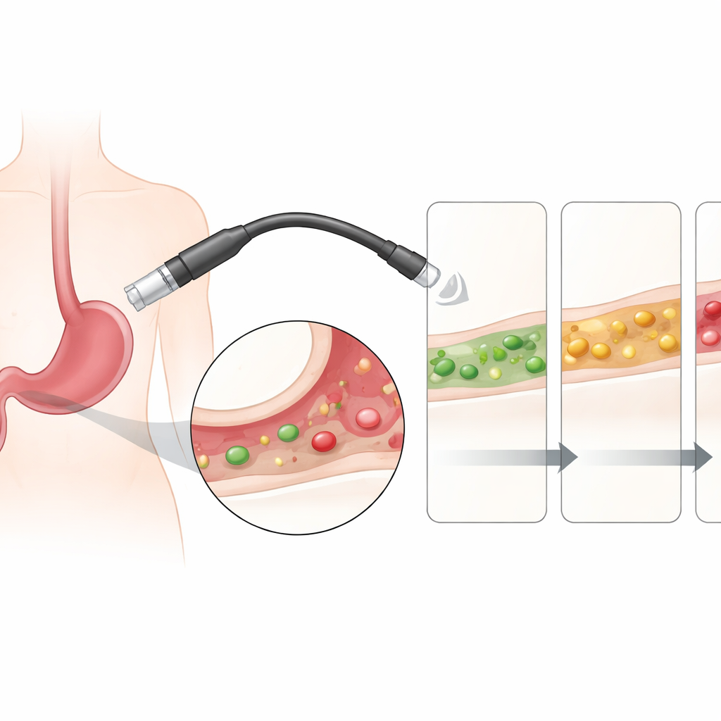
Peeking at lumps from inside the stomach
The authors focused on “small” stomach GISTs, defined as less than 2 centimeters across. Even though these are tiny, past data show that a meaningful portion already have spread or carry a higher risk of death. Current guidelines often recommend removing proven GISTs regardless of size, but the challenge is telling them apart from look‑alike benign lumps, such as muscle knots or fatty growths, without always cutting or sticking a needle into them. Endoscopic ultrasound (EUS) – a technique that combines endoscopy with ultrasound at the scope’s tip – can show where a lump sits within the stomach wall and how it looks inside, but on its own it is only about 70% accurate, especially for very small lesions.
Building a simple score from thousands of cases
To sharpen this blurred picture, the researchers retrospectively analyzed 1303 patients from several hospitals in China who had stomach wall lumps under 2 centimeters that were later removed and examined under the microscope. They split these patients into three groups: a training set to build the score, and two independent sets to check how well it works. For each lump, they recorded the patient’s basic information and detailed EUS features such as where in the stomach it sat, which layer of the wall it arose from, how bright or dark it looked on ultrasound, and whether it bulged inward or outward.
Four visual clues that matter most
Statistical testing showed that only four ultrasound‑based clues reliably marked a small lump as a GIST rather than some other type of tumor. These were: location in the upper stomach (cardia or fundus), origin in the deep muscle layer of the wall, a darker‑than‑surrounding (hypoechoic) appearance on ultrasound, and a tendency to grow outward from the stomach rather than just into its hollow center. The team turned these clues into a point system: 1 point for being in the upper stomach, 2 for arising from the muscle layer, 1 for being dark on ultrasound, and 2 for outward growth. Adding these up yielded a total score from 0 to 6, with higher values signaling a higher chance of GIST.
How well the score sorts risk
When tested, the score showed that as the total climbed, the proportion of true GISTs rose sharply. In the main development group, only about 8–10% of lumps with a score near 0–1 were GISTs, but more than 95% with scores of 5–6 were GISTs. In both the internal and external validation groups, the tool separated low‑, middle‑, and high‑risk lesions in a similar way. Overall performance, measured by how well the score distinguished GIST from non‑GIST, was strong, and the system proved especially good at ruling out disease: among patients labeled low‑risk, most indeed did not have GISTs. This means the score could help doctors feel more confident in simply watching some small lumps instead of rushing to biopsy or surgery.
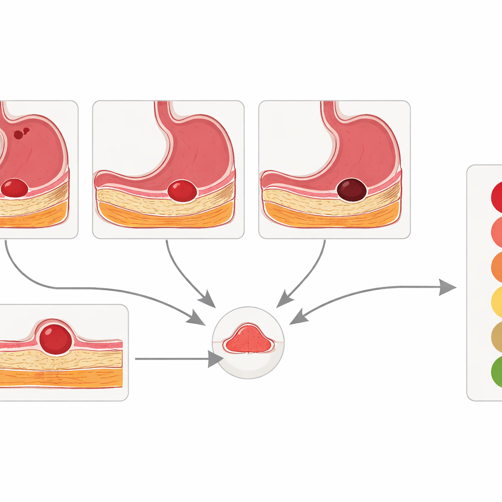
What this could mean for patients
The authors argue that their four‑point EUS‑based score can be easily folded into existing care pathways. After a routine EUS exam, a doctor could quickly total the points. Low scores (0–2) might support a “wait‑and‑see” approach with regular checkups; middle scores (3–4) could prompt more advanced tests, such as contrast‑enhanced ultrasound or needle sampling; and high scores (5–6) would favor timely removal of the lump. Although the study has limits – it is retrospective, from expert centers, and based on Chinese patients – it shows that careful reading of simple imaging features can meaningfully guide decisions. For patients, this could translate into fewer unnecessary procedures for benign lesions and more targeted treatment for those tiny stomach tumors that truly pose a threat.
Citation: Liu, L., Feng, Y., Zheng, S. et al. A novel risk-scoring system based on endoscopic ultrasound and clinical characteristics for the preoperative diagnosis of small gastric gastrointestinal stromal tumors. Sci Rep 16, 10279 (2026). https://doi.org/10.1038/s41598-026-41599-9
Keywords: gastric GIST, endoscopic ultrasound, risk score, submucosal tumors, early cancer detection