Clear Sky Science · en
Clinical accuracy of cephalometric analysis using deep learning–based automated landmark identification on CBCT in class I and class II malocclusions
Why smarter scans matter for your smile
When orthodontists plan braces or jaw surgery, they rely on precise measurements of the bones, teeth, and facial profile. Traditionally, experts trace these points by hand on X‑ray–like images, a task that is slow and prone to human variation. This study asks a simple but important question with real-world impact: can an artificial intelligence system accurately take over this measuring job in modern 3D scans, and if so, how safely can dentists trust its output when planning treatment?
From flat pictures to 3D head maps
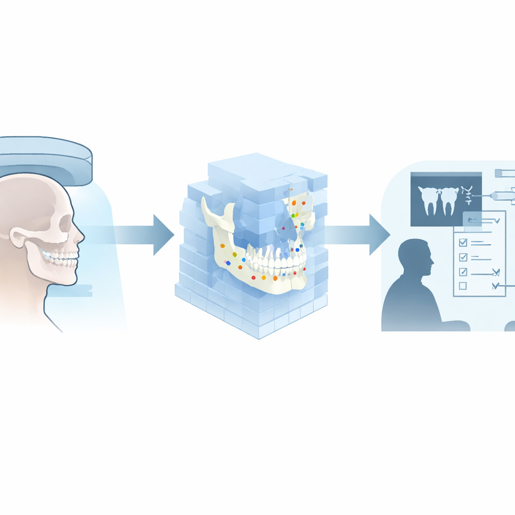
For decades, orthodontists used flat, two-dimensional images to measure jaw position and tooth angles. These images squeeze a 3D head into a single plane, which can hide subtle problems and distort distances. Cone-beam computed tomography (CBCT) now lets clinicians see the face and jaws in three dimensions, but carefully marking dozens of key points in 3D space is time-consuming and requires considerable training. Automated systems powered by deep learning promise to place these points—called landmarks—within fractions of a millimeter, potentially saving time and standardizing care.
Putting an AI measurer to the test
The research team built on an advanced deep learning model that had already been trained on nearly 500 CBCT scans. The algorithm was designed to spot 40 important bony, dental, and soft-tissue landmarks in 3D. For this study, the authors selected 75 new CBCT scans from patients with common bite types known as skeletal Class I and Class II malocclusions. Experienced orthodontists manually marked the landmarks on these scans; their work served as the “ground truth.” The AI then analyzed the same scans, and a custom software platform converted both sets of landmarks into 71 practical measurements—distances, angles, and ratios—that orthodontists actually use in diagnosis and treatment planning.
How close is close enough in the clinic?
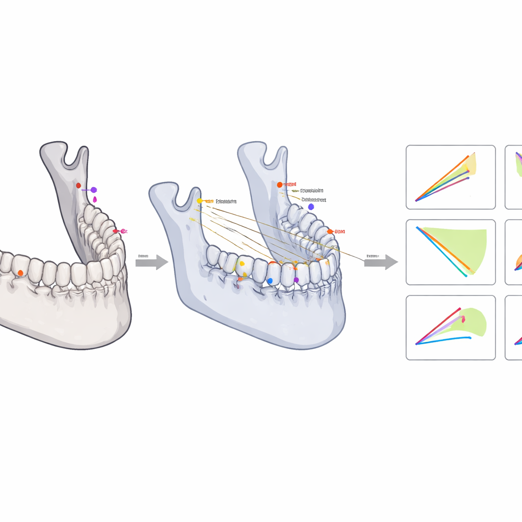
To judge the AI’s performance, the researchers compared its measurements with the expert-derived ones using several statistical tools. They looked at the average distance between corresponding landmarks, known as mean radial error, and at how often the AI landed within two millimeters of the expert marks. Overall, the AI’s landmarks were on average about 1.7 millimeters away from the experts’ points, and more than 60% fell within the tightest two-millimeter window. When these landmarks were translated into real clinical measurements, only 9 of 71 showed statistically significant differences—and even those differences were tiny, less than one millimeter or one degree, a range that orthodontists generally consider negligible in practice.
Where the robot ruler still struggles
The story becomes more nuanced when looking beyond simple averages. Using methods that assess not only the typical difference but also the spread of differences, the team found that nearly 60% of all AI-based measurements stayed comfortably within a clinically acceptable band of plus or minus two millimeters or degrees. Linear distances—such as the length of a jaw segment—were especially reliable when the underlying landmarks were located with better than two-millimeter precision. However, angle measurements, which depend strongly on the direction of small position errors, proved more sensitive. For some landmarks with fuzzy anatomical borders, such as the back corner of the jawbone (gonion) or the tips of developing incisor roots, the AI’s directional errors could noticeably affect angles, even when the average distance error looked acceptable.
A partnership between people and algorithms
For patients and clinicians, the key takeaway is that well-designed AI can already shoulder much of the heavy lifting in 3D orthodontic measurement, delivering results that closely match expert work for most clinically important distances and many angles. At the same time, the study shows that a single score describing average landmark error is not enough to guarantee trustworthy measurements, especially for angles and for landmarks in regions with blurry or variable anatomy. The authors conclude that automated 3D cephalometric analysis is ready to be used in routine care as a powerful assistant—but with a human in the loop. A brief manual check and fine-tuning of a few sensitive landmarks can turn the system into a reliable partner, helping orthodontists plan safer, more precise treatments while spending less time on tedious measurement chores.
Citation: Jiang, Y., AL-Mohana, R.A.A.M., Jiang, C. et al. Clinical accuracy of cephalometric analysis using deep learning–based automated landmark identification on CBCT in class I and class II malocclusions. Sci Rep 16, 10283 (2026). https://doi.org/10.1038/s41598-026-41408-3
Keywords: orthodontic imaging, 3D dental scans, artificial intelligence, automated measurements, CBCT