Clear Sky Science · en
A lightweight CNN for enhanced non-small cell lung cancer classification using CT scan image
Why faster lung scans matter
Lung cancer is one of the deadliest cancers, in part because it is often found late. Doctors increasingly rely on detailed CT scans and microscope images of lung tissue to spot early signs of disease, but reading these images takes time and expertise that are not always available, especially in smaller hospitals. This study introduces a compact artificial‑intelligence model, called MiniConvNet, designed to help doctors quickly and accurately distinguish between several major forms of non‑small cell lung cancer and healthy lungs, even on modest computers. 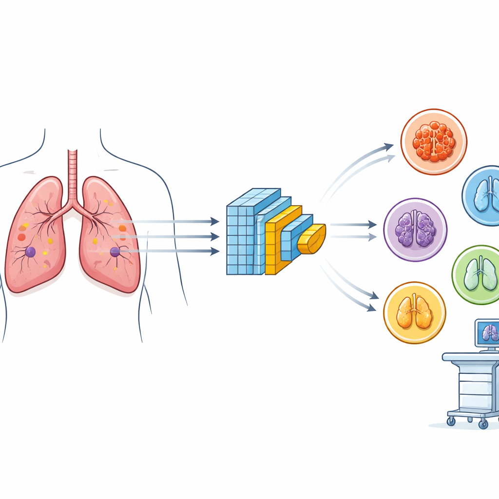
The challenge of spotting lung cancer early
Most lung cancer deaths are tied to non‑small cell lung cancer, an umbrella term that includes adenocarcinoma, squamous cell carcinoma, and large cell carcinoma. These subtypes behave differently, respond to different treatments, and can look quite similar on scans. Today, radiologists and pathologists examine CT scans and tissue slides to make these calls, but this work is labor‑intensive and can be especially difficult where there are few specialists. While modern deep‑learning systems can assist, many of the best‑performing models are large, slow, and require powerful graphics processors, making them hard to deploy in everyday clinics.
Building a smaller, smarter image reader
The authors set out to design a neural network that keeps the strengths of deep learning while trimming its bulk. MiniConvNet is a lightweight convolutional neural network, a type of algorithm well suited to analyzing images. It uses a series of small image filters, simple mathematical operations, and pooling layers that gradually condense visual information into a compact representation, followed by dense layers that make the final decision. With roughly half a million trainable parameters and a file size of only about 6 megabytes, MiniConvNet is far smaller than well‑known models such as VGG, ResNet, and Inception, which often contain tens of millions of parameters and require much more memory and processing power.
Putting the model to the test
To see how well their compact design worked, the researchers trained and evaluated MiniConvNet on two different kinds of lung images. The first was a CT scan collection of 900 images that included three cancer subtypes and healthy lungs. The second was a much larger set of 15,000 high‑resolution microscope images of lung tissue, covering similar classes. For both datasets, the images were resized to a standard format and underwent data augmentation—simple transformations such as flipping, rotation, and zooming—to help the model handle variations it might encounter in real practice. The same training settings, such as learning rate, batch size, and optimizer, were used for MiniConvNet and for a suite of popular reference models so that comparisons would be fair. 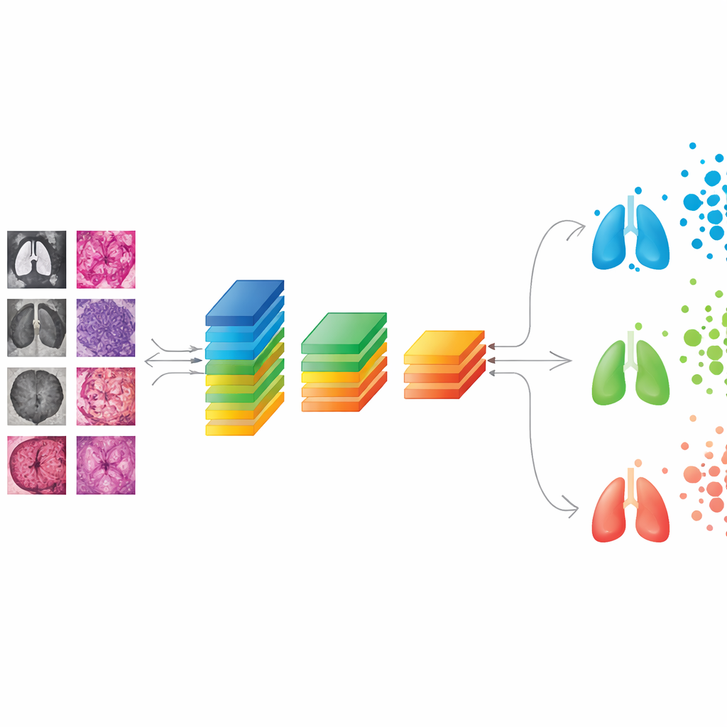
How the new model stacks up
On the CT scans, MiniConvNet achieved about 96% accuracy on the test set, clearly outperforming established models: the best of the larger networks, Inception V3, reached around 82% accuracy, while others such as VGG16, VGG19, ResNet‑50, EfficientNet, MobileNet, and ConvNeXt lagged behind. MiniConvNet also showed strong scores for precision, recall, and F1‑score across individual classes, indicating that it not only recognized cancer reliably but also distinguished between the three main subtypes and healthy lungs. On the tissue‑image dataset, the model again performed near the top, with test accuracy close to 97% and very balanced performance across categories. A five‑fold cross‑validation experiment—repeating training and testing on different splits of the data—confirmed that these results were stable rather than a lucky fluke.
Speed and practicality for real clinics
Beyond accuracy, the study emphasizes speed and resource use. MiniConvNet trained faster than many heavyweight models and delivered predictions in just a few milliseconds per image on standard hardware, while still handling both CT and tissue images well. These traits are crucial for point‑of‑care tools, where computers may be less powerful and doctors need rapid feedback. The researchers also showed that adding a regularization technique called dropout helped the model keep its performance high while reducing the risk of overfitting, an issue that often plagues deep learning on relatively small medical datasets.
What this means for patients and doctors
In everyday terms, the work suggests that a small, efficient AI program can help flag and categorize lung cancers with accuracy that rivals or surpasses much larger systems. MiniConvNet is compact enough to run on lower‑cost machines, making it a promising candidate for hospitals and clinics that currently lack advanced computing infrastructure. While larger and more varied patient datasets, as well as real‑world trials, are still needed before such a tool could be trusted in routine care, this study points toward a future in which fast, portable AI assistants support specialists in catching lung cancer earlier and tailoring treatment more precisely.
Citation: Baqir, M.A., Qayyum, S., Ashfaq, N. et al. A lightweight CNN for enhanced non-small cell lung cancer classification using CT scan image. Sci Rep 16, 12985 (2026). https://doi.org/10.1038/s41598-026-41401-w
Keywords: lung cancer, CT scan, deep learning, convolutional neural network, medical imaging