Clear Sky Science · en
Computational fluid dynamics-based flow field simulation and optimization of negative-pressure stone removal: stone size, position, and sheath geometry
Why this matters for people with kidney stones
Kidney stones are common and notoriously painful, and modern keyhole surgery can usually break them into tiny pieces. But getting those fragments out safely and completely is still a challenge. This study uses advanced computer simulations to peer inside a suction tube used during flexible kidney stone surgery, asking a practical question that matters to patients and surgeons alike: how can the shape of the tube, the suction strength, and the size and position of stone fragments be tuned so that more stones are cleared in one go, with fewer complications afterward? 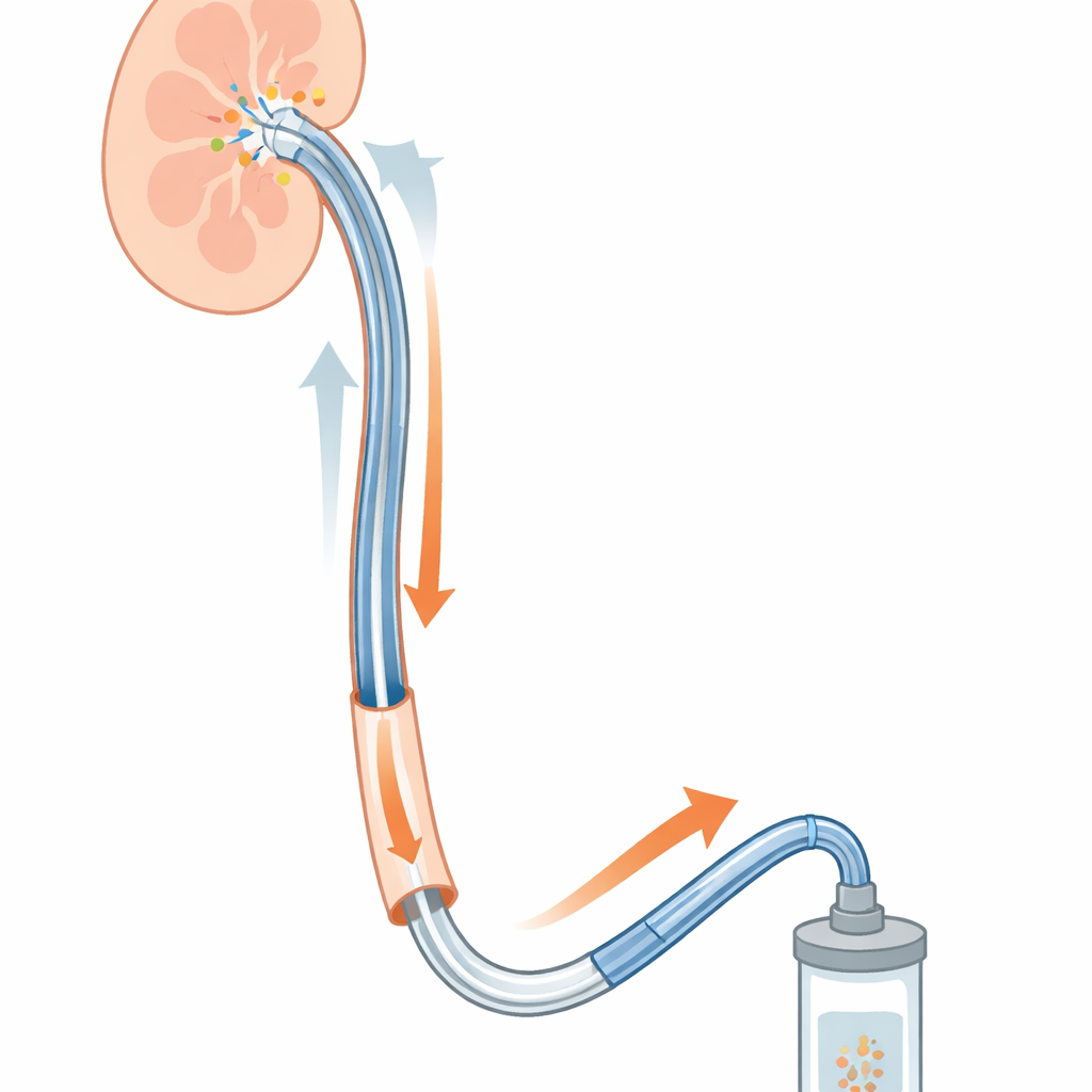
How stones are cleared with gentle suction
Many stone operations today use a thin flexible telescope passed through a hollow tube called a ureteral access sheath, which runs from the bladder up to the kidney. The telescope delivers laser energy to break the stone and also flushes water into the kidney, while the sheath connects to a suction source that draws water and fragments out. In practice, surgeons notice that some fragments are whisked away easily while others stubbornly remain or even seem to bounce around. Until now, these behaviors were mostly explained by experience and trial-and-error rather than by a detailed understanding of how fluid and fragments actually move inside the sheath.
Using virtual surgery to see the invisible
The researchers built a three-dimensional computer model that included the access sheath, the flexible scope, the urinary passage, and idealized spherical stone fragments between 1 and 3 millimeters wide. They simulated how water flows when it is pushed out of the scope tip and at the same time pulled back through the sheath by negative pressure. By changing stone size, suction strength, sheath diameter, and how far a stone sat from the scope tip, they could predict the forces acting on each fragment and whether it would be drawn toward the sheath opening or pushed away. This virtual approach let them explore complex flow patterns that would be very hard to measure directly in patients. 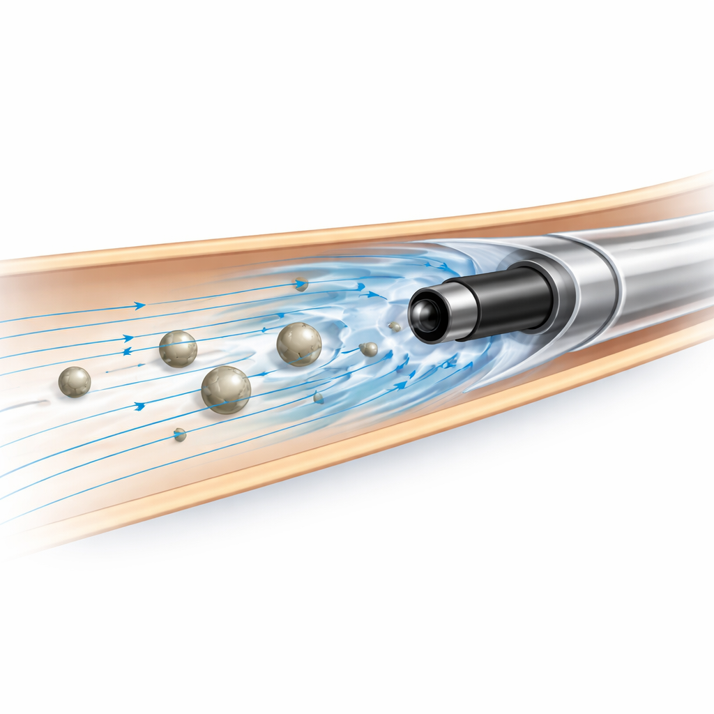
What stone size and position really do
The simulations showed that stone size and distance from the scope tip strongly shape how well suction works. Tiny 1 millimeter fragments felt their strongest pull when they were about 5 millimeters in front of the scope tip. Medium 2 millimeter fragments had a sweet spot much farther away, around 45 millimeters, and could actually be pushed away when they were extremely close to the tip, where the outgoing irrigation stream dominates. The largest 3 millimeter fragments experienced the greatest overall pulling force, peaking around 15 millimeters from the tip, but they also stirred up more chaotic flow, causing them to move in a jumpy, unstable way. Behind each stone, swirling low-pressure zones formed that could help nudge fragments along but also make their paths less predictable.
The "high-efficiency" zone inside the body
By comparing many combinations, the team identified a practical working window just a few millimeters long, roughly 5 to 15 millimeters in front of the scope tip, where suction-driven stone transport is most reliable. Within this zone, the flow tends to be more orderly and the pressure differences across a stone are well aligned to pull fragments into the sheath. Outside this range, especially extremely close to the tip or far upstream, irrigation flow, turbulence, and swirling vortices can oppose or destabilize stone motion. The simulations also suggested that a commonly used sheath size (12/14 French) offers a good balance: large enough to clear fragments efficiently but not so large that the flow becomes wildly unstable or potentially unsafe for surrounding tissue.
What this means for future stone treatments
For patients, the work does not change today’s operating room rules overnight, but it offers a scientific basis for improving them. The study suggests that surgeons could improve stone-free rates by adjusting where they break and position fragments so that they are drawn into the high-efficiency zone, rather than sitting directly at the scope tip or far away. It also points the way toward smarter sheath designs and suction systems that adapt to different stone sizes. While the model simplifies the real anatomy and movement inside the body, it provides a roadmap for future tools and guidelines that could make stone surgery safer, faster, and more likely to leave patients truly stone-free after a single procedure.
Citation: Tian, C., Liu, J., Di, Q. et al. Computational fluid dynamics-based flow field simulation and optimization of negative-pressure stone removal: stone size, position, and sheath geometry. Sci Rep 16, 11265 (2026). https://doi.org/10.1038/s41598-026-41399-1
Keywords: kidney stones, flexible ureteroscopy, negative pressure suction, computational fluid dynamics, ureteral access sheath design