Clear Sky Science · en
Reliability and agreement study of three-dimensional measurement for femoral head displacement indicators after femoral neck fractures
Why hip fracture alignment matters
When an older adult breaks the narrow "neck" of the thigh bone just below the hip, surgeons rush to realign and fix the bone with screws. Whether that repair is truly well aligned can shape the person’s future mobility, pain level, and even the risk that the ball of the hip joint slowly dies and collapses. This study asks a practical but crucial question: can modern three-dimensional (3D) CT scans provide hip surgeons with precise, trustworthy measurements of how well the broken bone has been put back into place?
From flat X-rays to 3D pictures
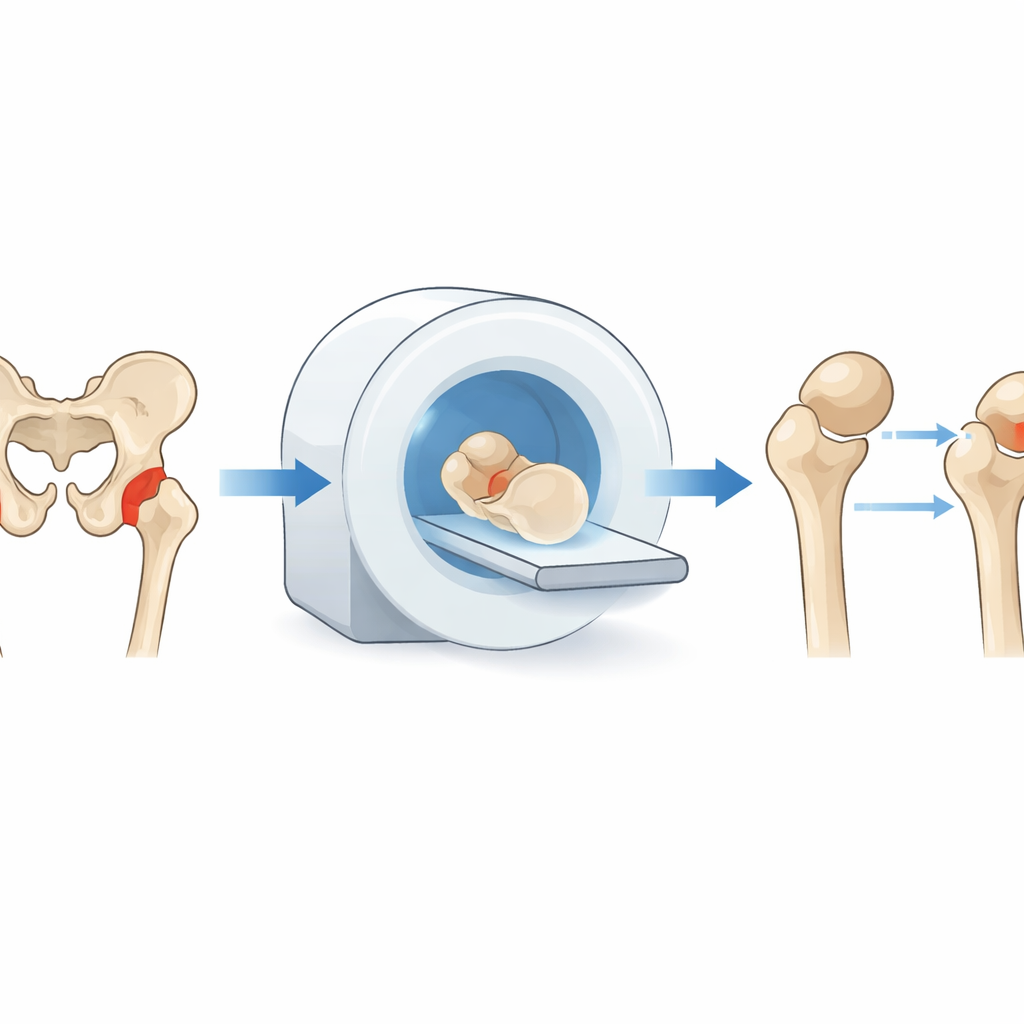
Traditionally, doctors judge hip fracture severity and the quality of surgical repair using standard two-dimensional X-rays. These flat images can be misleading. Bones overlap, patient positioning varies, and a repair that looks perfect in one view may still be slightly off in real 3D space. Earlier work using CT-based 3D reconstructions revealed that even so-called "non-displaced" fractures often show meaningful shifts in the position and rotation of the femoral head — the ball at the top of the thigh bone. Because these subtle shifts are linked to complications like poor healing and bone death, there is growing interest in using 3D measurements to capture them more accurately.
How the researchers measured bone shifts
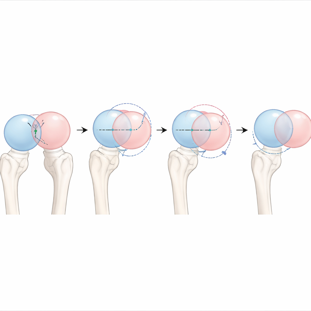
The team analyzed CT scans taken before and after surgery in 100 patients with femoral neck fractures treated using cannulated screws, a common internal fixation method. Using specialized software, they built 3D models of both hips for each patient. Assuming that left and right hips are normally nearly mirror images, they flipped the healthy side in the computer and carefully aligned it with the broken side. They then calculated how far the injured femoral head had shifted compared with its expected normal position, using three key indicators: the displacement of a small pit on the head of the femur, the shift of the center of the ball itself, and the overall 3D angle that describes how the head is twisted or tilted in space.
Testing if experts see the same thing
To find out how dependable these measurements are, three experienced observers independently repeated the entire workflow on the same set of scans. The authors then used several statistical tools to judge how closely the observers agreed. Before surgery, all three measures of displacement showed large, consistent differences between the injured and healthy sides, and the agreement between observers was very high: their numbers for the same patient were very close to one another. After surgery, the femoral heads were much closer to their normal positions, as expected from successful reduction and fixation. Agreement remained acceptable but became more modest, especially for angles measured when projecting the 3D geometry onto the side-view (sagittal) plane, where anatomical landmarks are less distinct and small choices in how to view the bone can shift the result.
Why agreement seems lower after good surgery
At first glance, it might seem worrying that statistical measures of reliability were lower after surgery than before. The authors explain that this is partly a mathematical illusion. Once fractures are well reduced, patients’ measurements cluster into a narrow, near-normal range. In this situation, even tiny differences between observers can make correlation-based statistics look weaker, without reflecting a real loss in precision. Other analyses, such as Bland–Altman plots that examine actual differences in millimeters or degrees, showed that most readings still fell in a narrow, clinically acceptable band. The main weak spot was again the sagittal plane, where aligning 3D models and defining a standard view is technically challenging and prone to variation.
What this means for patients and future tools
For patients, this work reinforces the idea that detailed 3D imaging can help doctors understand how badly a hip fracture has shifted and how well it has been corrected, factors that are tied to the risk of long-term problems. For researchers and clinicians developing new guidelines, the study provides evidence that CT-based 3D measurements of hip fracture displacement are highly reliable before surgery and generally dependable after surgery, provided their limitations are recognized. The authors conclude that future progress will depend on more standardized ways of aligning bone models in all three anatomical planes and on semi-automated software that reduces human judgment calls. With those refinements, 3D reconstruction could become a routine, objective tool to guide treatment and predict outcomes for people with hip fractures.
Citation: Cui, S., Yu, J., Zhao, L. et al. Reliability and agreement study of three-dimensional measurement for femoral head displacement indicators after femoral neck fractures. Sci Rep 16, 11303 (2026). https://doi.org/10.1038/s41598-026-41210-1
Keywords: hip fracture, femoral neck, 3D CT, bone alignment, surgical outcomes