Clear Sky Science · en
SARS-CoV-2 nucleocapsid protein-specific monoclonal antibodies as tools for studying its antigenic structure and interaction with host cells
Why this research matters
Even as vaccines and treatments have softened the blow of COVID-19, the virus that causes it, SARS-CoV-2, continues to evolve. Most attention has focused on the virus’s outer spike protein, which changes rapidly. This study turns the spotlight on a different, more stable part of the virus—the nucleocapsid protein inside the viral shell—and creates precise antibody tools to better detect the virus and probe how it interacts with our cells and immune system.
A hidden but crucial viral part
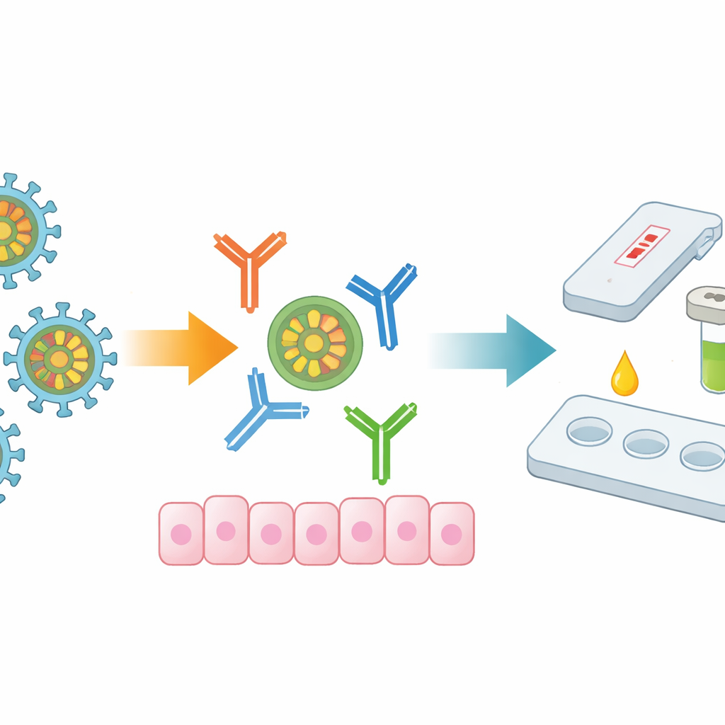
Inside every coronavirus particle sits the nucleocapsid protein, which wraps and organizes the viral genetic material. Because this protein changes far less than the spike, it looks very similar across many SARS-CoV-2 variants and even to the virus that caused the 2002–2004 SARS outbreak. It is also produced in large amounts during infection and strongly stimulates antibody responses, even in people with mild or symptom-free disease. These qualities make the nucleocapsid an attractive target both for diagnostic tests and for understanding how the virus disrupts the body’s defenses and drives inflammation.
Building precise antibody tools
The researchers immunized mice with either the full nucleocapsid protein or a shortened version missing part of its beginning section, then fused the animals’ immune cells with tumor cells to create hybridoma cell lines that continuously produce specific antibodies. They isolated nine monoclonal antibodies that recognize SARS-CoV-2 nucleocapsid with very high binding strength. Using a suite of lab techniques, they confirmed that all nine antibodies recognize the natural viral protein inside infected cells and also bind nucleocapsid from the Omicron variant, which carries multiple changes in other parts of its genome.
Pinpointing the protein’s “hot spots”
To learn exactly where on the nucleocapsid these antibodies bind, the team engineered overlapping fragments of the protein and tested which fragments each antibody recognized. This mapping showed that most antibodies target regions involved in key jobs: binding viral RNA, pairing nucleocapsid molecules together, and helping the protein form droplets that regulate viral replication and immune signaling. Eight of the nine antibodies were found to latch onto such functionally active zones, and their target sequences remain almost unchanged across major variants, including recent Omicron offshoots. Interestingly, when the scientists compared these mouse antibody targets with those typically recognized by human antibodies from people who had recovered from COVID-19, there was little direct competition, suggesting that these monoclonal antibodies highlight complementary, less commonly targeted regions of the protein.
Watching the protein enter cells
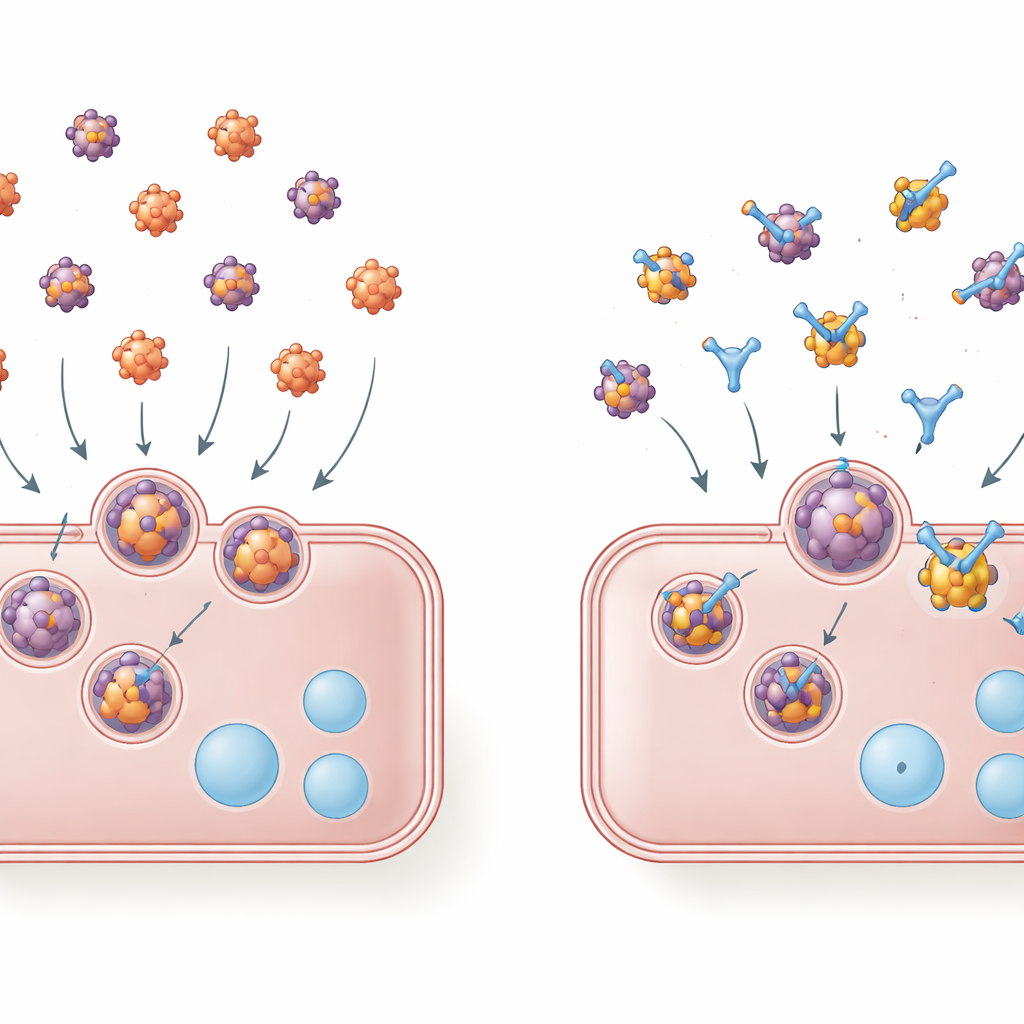
Beyond its role inside infected cells, nucleocapsid can also appear outside cells, where it sticks to cell surfaces and is taken up by a process similar to receptor-mediated endocytosis. This external protein may help trigger harmful inflammation. The authors labeled Omicron nucleocapsid with a fluorescent dye and exposed human lung cells to it. Under the microscope, they saw the labeled protein move into the cells and collect in compartments resembling lysosomes, a pattern that was strongly reduced when they blocked endocytosis with a chemical inhibitor. This confirmed that free nucleocapsid protein can indeed be pulled into lung cells through an active uptake pathway.
Antibodies that slow cellular entry and aid detection
The team then tested whether their monoclonal antibodies could interfere with this uptake. They pre-mixed fluorescent nucleocapsid with each antibody before adding it to lung cells. Several antibodies, notably those named 4B3, 7F10, 16D9, and 18A8, reduced how much nucleocapsid entered the cells, as judged by lower fluorescence inside the cells. Others, which bind nearby or different regions, did not have this blocking effect. Separately, the researchers combined selected antibodies into a “capture and detection” pair and built a sandwich-style lab test. One antibody anchored nucleocapsid to a surface, while a second, enzyme-linked antibody generated a color signal. This setup detected both original and Omicron nucleocapsid over a wide range of concentrations, highlighting the antibodies’ promise for future diagnostic assays.
What the findings mean
By creating and precisely characterizing nine monoclonal antibodies that latch onto crucial, conserved regions of the SARS-CoV-2 nucleocapsid protein, this study delivers versatile tools for COVID-19 research. These antibodies reliably spot the viral protein in infected cells, help build sensitive nucleocapsid-based tests that should remain useful across emerging variants, and, in some cases, physically hinder the protein from entering lung cells. For non-specialists, the key message is that looking beyond the famous spike protein to this more stable inner protein can improve diagnostics and deepen our understanding of how the virus manipulates our cells and immune systems, opening doors to new ways of detecting and perhaps one day disarming the virus.
Citation: Rimkutė, A., Simanavičius, M., Dalgėdienė, I. et al. SARS-CoV-2 nucleocapsid protein-specific monoclonal antibodies as tools for studying its antigenic structure and interaction with host cells. Sci Rep 16, 11461 (2026). https://doi.org/10.1038/s41598-026-40984-8
Keywords: SARS-CoV-2 nucleocapsid, monoclonal antibodies, COVID-19 diagnostics, viral pathogenesis, host–virus interaction