Clear Sky Science · en
Plasma lipid remodeling during the early recovery phase after myocardial infarction
Why heart attack recovery is more than feeling better
When someone survives a heart attack, doctors often focus on opening clogged arteries and prescribing medicines to lower cholesterol and calm inflammation. Yet inside the bloodstream, far more subtle changes are unfolding. This study peeks under the hood during the first month after a heart attack and shows that the fats circulating in the blood — not just cholesterol, but dozens of specialized lipids — remain disturbed even when patients already feel well. These hidden shifts could help explain why some hearts heal smoothly while others drift toward heart failure or another cardiac event.
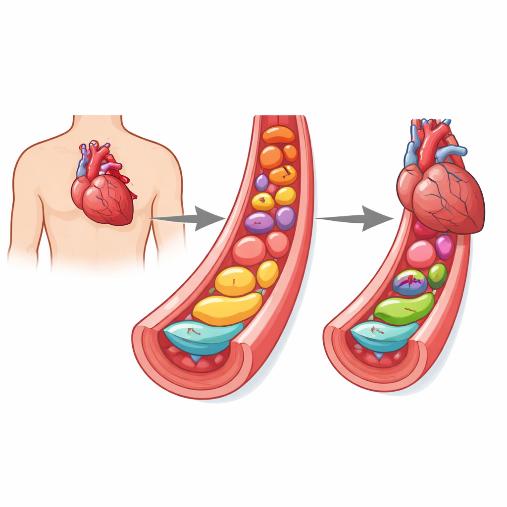
The hidden players: special fats in the blood
The researchers focused on complex fats that help build cell membranes and send danger or repair signals in the body. These included phospholipids (key building blocks of cell walls), plasmalogens (a sub‑group that act like built‑in antioxidants), and sphingolipids such as sphingomyelins and ceramides, which are involved in cell stress and death. Rather than just measuring total cholesterol, the team used advanced mass spectrometry to profile more than a hundred individual lipid species in blood plasma from three groups: people in the first day after a heart attack, the same condition 3–6 weeks later during planned readmission, and healthy volunteers of similar age and sex.
What happens to lipids during a heart attack
In the acute phase of a heart attack, the blood lipid landscape changed dramatically. Many phospholipids rich in polyunsaturated fatty acids — especially certain phosphatidylcholines and phosphatidylethanolamines — were sharply depleted. These molecules are especially fragile in the face of the burst of reactive oxygen species that accompanies the sudden loss and restoration of blood flow, so their loss likely reflects oxidative damage to cell membranes. At the same time, simpler breakdown products called lysophospholipids and several sphingomyelins rose, consistent with inflammation and membrane injury. Using statistical pattern analysis, the researchers showed that the overall lipid profile of heart‑attack patients was clearly distinct from that of healthy people.
The early recovery window: better, but not back to normal
Three to six weeks later, when patients were clinically stable and on modern drug therapy, their blood lipids told a more complicated story. Some lipid species moved back toward normal: inflammatory lysophospholipids and several sphingomyelins dropped to levels similar to healthy controls, and certain saturated phosphatidylcholines rebounded. A subset of plasmalogens derived from phosphatidylcholine also partially recovered, hinting that antioxidant defenses and membrane repair were being rebuilt. However, other protective lipids — especially plasmalogens linked to phosphatidylethanolamine — remained depressed, suggesting that oxidative and metabolic stress were far from fully resolved. Overall, the recovery group’s lipid pattern sat between that of acute patients and healthy controls, signaling a biochemical state of “in‑between” healing.
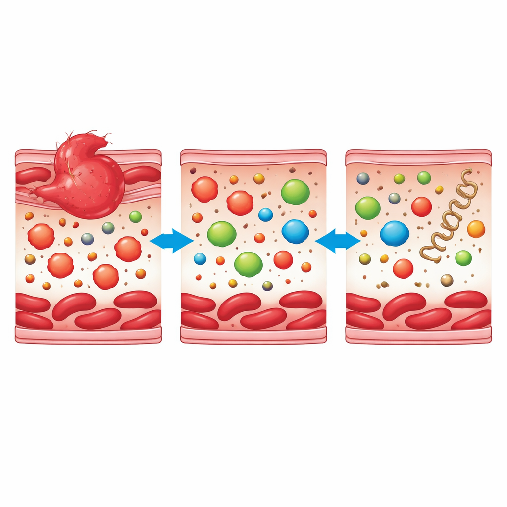
Stubborn warning signals that linger
Not all lipid changes faded with time. Ceramides, a class of sphingolipids known to promote cell death, inflammation, and harmful remodeling of heart tissue, stayed high in both the acute and early recovery patients compared with healthy volunteers. This persistent ceramide signature points to ongoing stress in the heart and blood vessels even when symptoms have settled and standard tests look reassuring. Pathway analysis reinforced this picture: networks related to membrane phospholipids, ether lipids (which include plasmalogens), and sphingolipids were strongly disrupted during the heart attack and remained partially disturbed weeks later, despite aggressive use of statins and other guideline‑directed drugs.
What this means for patients and future care
To a layperson, the message is that “normal” recovery from a heart attack is more than closing an artery or lowering bad cholesterol. Deep inside the bloodstream, the mix of structural and signaling fats remains altered for weeks, with some protective lipids still low and some harmful ones, like ceramides, stubbornly high. This work does not yet change day‑to‑day treatment, but it suggests that detailed lipid fingerprints could one day help identify patients whose hearts are silently struggling to heal — long before problems show up on scans or symptom checklists. With larger, longer studies, such lipid markers might guide more personalized follow‑up and therapies aimed not only at arteries, but also at restoring the body’s microscopic repair chemistry.
Citation: Myszko, M., Bychowski, J., Radziwon, P. et al. Plasma lipid remodeling during the early recovery phase after myocardial infarction. Sci Rep 16, 9916 (2026). https://doi.org/10.1038/s41598-026-40864-1
Keywords: myocardial infarction, lipidomics, plasmalogens, ceramides, cardiac remodeling