Clear Sky Science · en
Sleep disorders and structural alterations in brain regions linked with motivation: a neuroimaging meta-analysis
Why broken sleep matters for everyday life
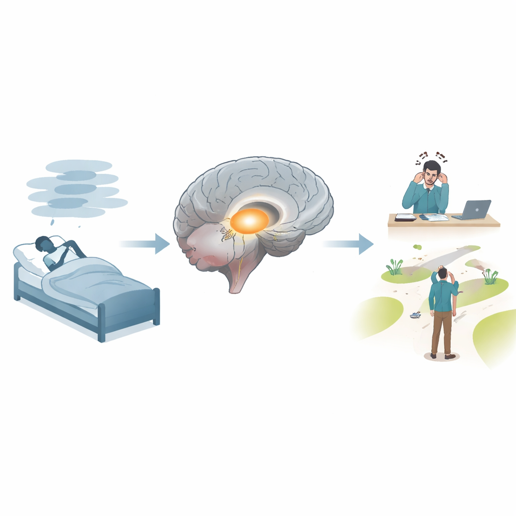
Poor sleep is more than just feeling tired. People with chronic sleep disorders are more likely to make mistakes at work, have emotional blowups, take unnecessary risks, and get into accidents. These problems cost society billions of dollars each year. Yet we still know surprisingly little about how long‑term sleep disruption physically reshapes the brain, especially in regions that help us stay focused, control our emotions, and pursue goals. This study pulled together dozens of brain‑scan experiments to ask a simple but powerful question: are there common weak points in the brains of people with different kinds of sleep disorders, and do those weak points sit in circuits that drive motivation and decision‑making?
Two broad kinds of troubled sleep
Sleep disorders come in many forms, but this work focused on two big families. One group, called parasomnias, includes conditions like sleepwalking, sleep terrors, nightmare disorder, and rapid eye movement (REM) sleep behavior disorder, where people act out dreams or show strange behaviors during sleep. The other group, dyssomnias, covers problems with falling or staying asleep or staying awake, such as insomnia, narcolepsy, restless legs syndrome, and breathing‑related disorders like obstructive sleep apnea. Although both types lead to unrefreshing sleep and daytime struggles, they may stem from different breakdowns in the brain’s control of arousal, internal awareness, and reward.
Scanning the literature instead of single brains
Instead of running a new experiment with a small group of volunteers, the researchers carried out a large meta‑analysis: a statistical "study of studies." They searched the medical literature up to late 2025 and selected 57 brain‑imaging papers that measured differences in brain structure between adults with a diagnosed sleep disorder and healthy sleepers. All studies reported precise three‑dimensional coordinates showing where brain tissue was thinner or thicker. Using a method called Activation Likelihood Estimation, the team treated each coordinate as a probability cloud and looked for spots where many papers independently pointed to the same region. They did this first by pooling all sleep disorders together, then by analyzing parasomnias and dyssomnias separately.
A shared hub for attention goes offline
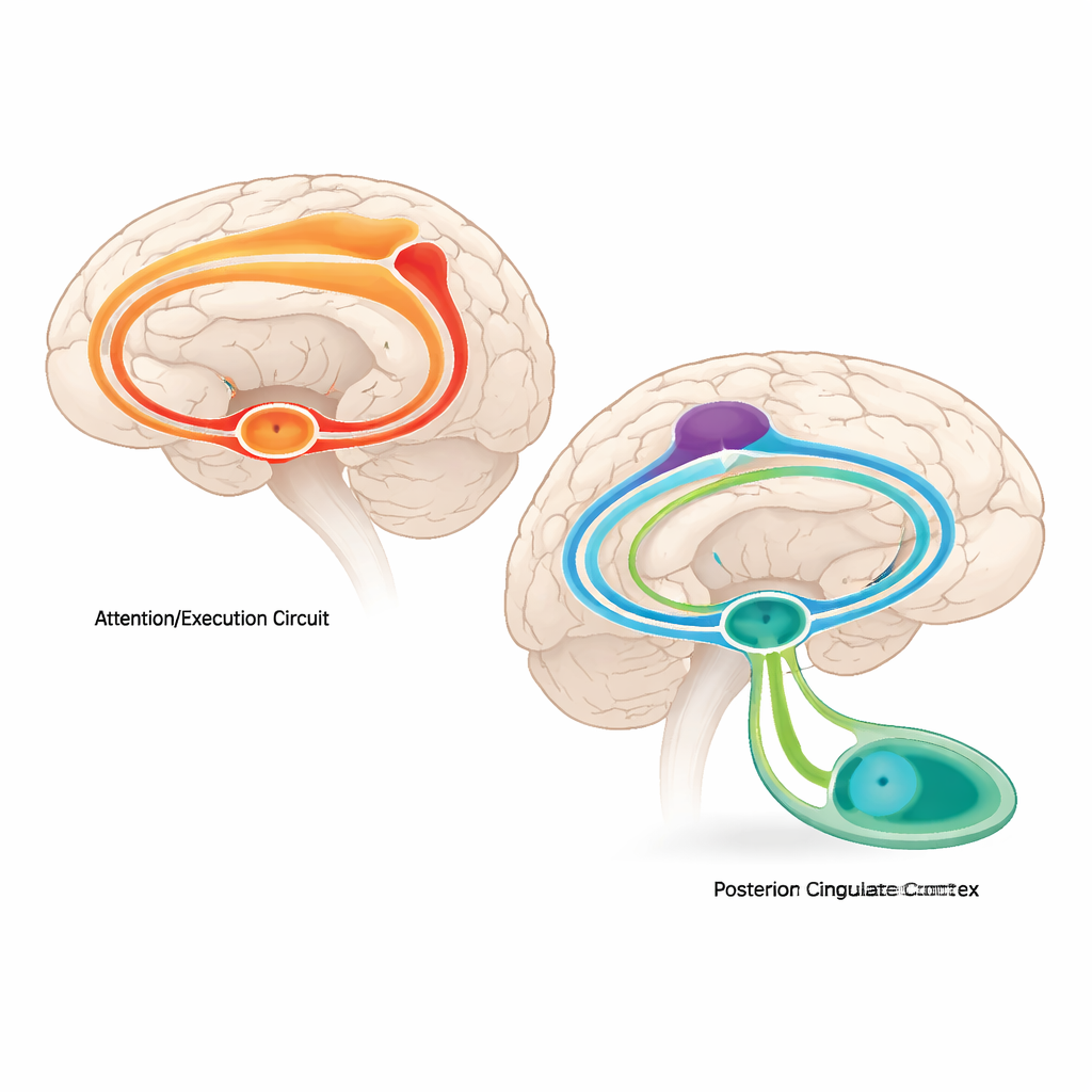
Across all types of sleep disorders combined, one brain region stood out: the thalamus, specifically a subdivision called the pulvinar. This deep structure acts as a relay and gatekeeper for sensory information, helping the cortex focus on what matters and ignore distractions. People with sleep disorders tended to have less gray matter in this area, suggesting subtle loss or thinning of tissue. When the authors examined large databases of functional brain scans from many different tasks, they found that this thalamic zone normally works as part of a wide‑ranging network with frontal and parietal regions that supports paying attention, switching tasks, and monitoring performance. Damaging or weakening this hub, they argue, could help explain why sleep‑deprived or sleep‑disordered individuals are more prone to lapses, slower responses, and everyday errors.
When sleepwalking meets the brain’s inner compass
The picture looked different for parasomnias. In these disorders, the only consistent structural change was a loss of gray matter in the posterior cingulate cortex, a region on the midline near the back of the brain. This area is a key node in the so‑called default mode network, which supports self‑reflection, daydreaming, and the internal sense of "me." By tracing its typical partners in large databases, the researchers showed that the posterior cingulate usually works closely with frontal areas that track value and with deep reward regions like the striatum and insula. Together, this network helps weigh options, assign importance to outcomes, and steer motivated behavior. Structural weakening here could make it harder to smoothly integrate feelings, memories, and goals—potentially contributing to bizarre or poorly controlled actions that emerge during parasomnia episodes.
Different sleep problems, different brain networks
Interestingly, when the team tried to look for a single structural pattern that defined dyssomnias alone, they did not find any brain region that consistently shrank or grew across studies. The authors suggest that this may reflect the sheer diversity of conditions lumped into this category and the still‑limited number of high‑quality imaging studies for each one. Even so, the broader pattern hints at an important theme: while all sleep disorders seem to tap into attention‑related circuits centered on the thalamus, parasomnias additionally involve the brain’s valuation and self‑monitoring systems centered on the posterior cingulate. In other words, different kinds of troubled sleep may tilt different large‑scale networks off balance.
What this means for people struggling with sleep
For non‑specialists, the takeaway is that chronic sleep disorders are not just about feeling drowsy—they are tied to measurable changes in brain hubs that support focus, motivation, and wise choices. Thalamic changes may leave people more vulnerable to distraction and mistakes, while posterior cingulate changes in parasomnias may warp how the brain assigns value and keeps track of one’s own actions, even during sleep. Recognizing that these conditions disturb distinct yet overlapping brain networks could help clinicians design more targeted treatments, from behavioral strategies that protect attention to therapies that stabilize reward and motivation systems. Better sleep, in this view, is not only about more hours in bed but about restoring the brain circuits that keep us alert, steady, and goal‑directed during the day.
Citation: Crooks, K.E., Hampson, C.L., Peraza, J.A. et al. Sleep disorders and structural alterations in brain regions linked with motivation: a neuroimaging meta-analysis. Sci Rep 16, 11130 (2026). https://doi.org/10.1038/s41598-026-40818-7
Keywords: sleep disorders, brain networks, motivation, thalamus, parasomnias