Clear Sky Science · en
A scanning electron microscopy—based approach to explore subpodocyte space remodeling in diabetic kidneys of mice and humans
Why tiny kidney spaces matter
Diabetes is well known for damaging the kidneys, but much of that harm begins in microscopic corners that ordinary microscopes cannot easily see. One such nook, called the subpodocyte space, sits beneath the cells that wrap around the kidney’s filtering loops and may be an early warning sign of trouble. This study introduces a faster, more practical way to picture these hidden spaces in both mice and people, bringing laboratory insights closer to real-world diagnosis and treatment of diabetic kidney disease.
Looking closer at the kidney’s hidden filters
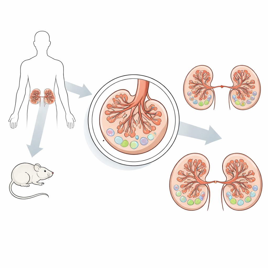
The filters inside each kidney are made of tiny loops of blood vessels wrapped by specialized cells that help keep useful proteins in the blood while letting waste pass into urine. Between these surface cells and the supporting layer beneath them lies a very narrow compartment, the subpodocyte space, that shapes how fluid moves through the filter. Earlier work in animals suggested that this space swells in diabetes, putting extra strain on the filter and contributing to kidney damage. However, studying this region in detail required a demanding technique called transmission electron microscopy, which is slow, covers only small areas, and is hard to apply to routine patient biopsies.
Adapting a sharper microscope for real-world samples
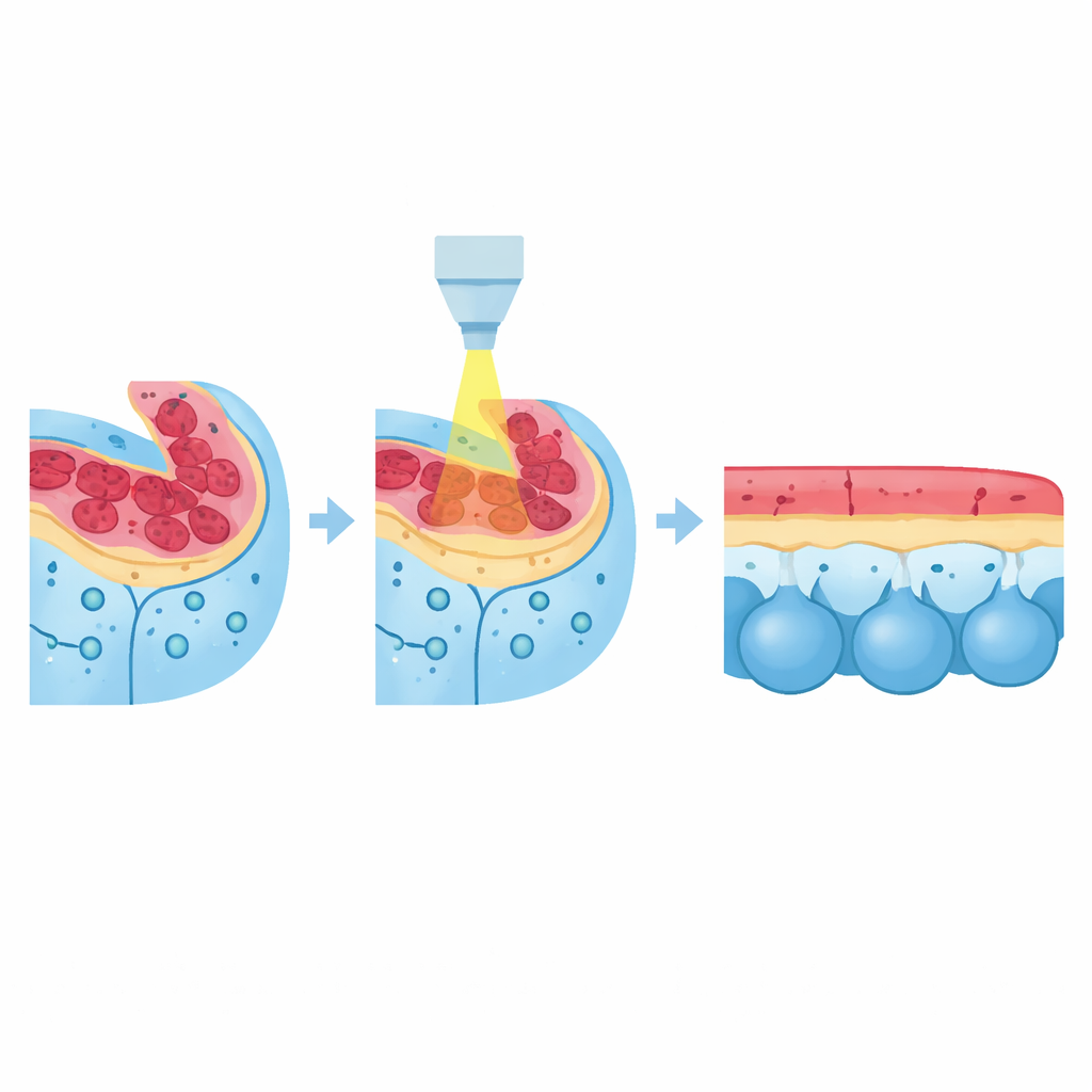
The authors had previously developed a scanning electron microscopy protocol that could capture near–electron-microscope detail over wider areas, but it depended on flushing the blood vessels with fixative, a step that is simply not possible when a doctor takes a kidney biopsy from a patient. In the new work, they re-engineered this approach so that it works on non-perfused tissue—the type actually obtained in clinics—where red blood cells and uneven electrical properties can distort images. By adjusting the energy of the electron beam and turning off a high-current mode, they reduced electrical “charging” that causes blurring and streaks, and achieved stable, high-resolution images of semi-thin kidney sections without extra coatings or elaborate preparation.
What the new method reveals in diabetic mice
With this optimized setup, the team compared healthy and diabetic mice, examining kidneys that had been perfused and those that had not. In all conditions, diabetic animals showed clear enlargement of the subpodocyte space, with broader pockets forming beneath the filtering cells. Careful measurements confirmed that, even without perfusion, diabetic mice had a larger subpodocyte area overall, a greater fraction of each filtering tuft occupied by this space, and more subpodocyte area per filtering cell than healthy controls. At the same time, diabetic mice had enlarged filtering units and fewer of these key cells per unit area, signs of structural overload and wear that are typical of diabetic kidney disease. Together, these observations show that the method can reliably capture meaningful disease-related changes under realistic tissue conditions.
Seeing early changes in human kidney biopsies
Encouraged by the mouse results, the researchers applied the protocol to human kidney samples: one from a person without kidney disease and one from a person with early diabetic kidney damage. They were able to image entire cross-sections of the kidney filters at high resolution, something that would be far more time-consuming with traditional electron microscopy. Using the same measurement approach as in mice, they found that the subpodocyte space occupied a much larger area in the diabetic patient’s glomeruli than in the control. The technique also revealed subtle signatures of injury, such as fragmentation of the filtering cells, all while saving time and preserving tissue for possible follow-up studies.
What this means for people with diabetes
For non-specialists, the key message is that this study provides a practical new way to see where diabetic kidney damage begins—inside a narrow, previously hard-to-access space beneath the kidney’s filtering cells—using tissue prepared in the same way as ordinary biopsies. By showing that the subpodocyte space is measurably enlarged in both diabetic mice and a human patient, even at an early stage of disease, the work supports its role as an early structural warning sign. The improved imaging method could speed up detailed kidney assessments, help researchers track how diabetes reshapes the kidney filter over time, and eventually aid clinicians in identifying and monitoring kidney injury before irreversible scarring sets in.
Citation: Conti, S., Benigni, A., Remuzzi, G. et al. A scanning electron microscopy—based approach to explore subpodocyte space remodeling in diabetic kidneys of mice and humans. Sci Rep 16, 10095 (2026). https://doi.org/10.1038/s41598-026-40816-9
Keywords: diabetic kidney disease, electron microscopy, podocyte injury, kidney biopsy, glomerular filtration