Clear Sky Science · en
Liquid–liquid phase separation by caged coacervating peptides
Light as a Gentle Switch for Tiny Droplets
Many of the busy reactions inside our cells take place in tiny liquid droplets that have no surrounding membrane. These droplets behave a bit like oil droplets in a salad dressing, concentrating certain molecules while excluding others. The study described here shows how researchers built an artificial version of such droplets from short protein fragments, and then used blue light as a gentle on–off switch to assemble and partially dissolve them. This light-controlled system could one day help deliver drugs, regulate reactions in test tubes, or shed light on how similar droplets work in living cells.
How Nature Uses Droplets Without Membranes
Cells organize their contents not only with classic membrane-bound compartments such as the nucleus, but also with softer, liquid droplets that form when biomolecules separate into two coexisting liquid phases. This behavior, called liquid–liquid phase separation, underlies structures like stress granules and nucleoli. It can be helpful, for example by speeding up reactions, but it is also linked to diseases when it goes wrong, contributing to harmful protein clumps in Alzheimer’s and Parkinson’s disease. To understand and eventually control these processes, scientists are building simplified droplet systems from designed molecules like peptides and nucleic acids that can mimic cellular droplets in a controlled way.
Designing Droplets That Respond to Light
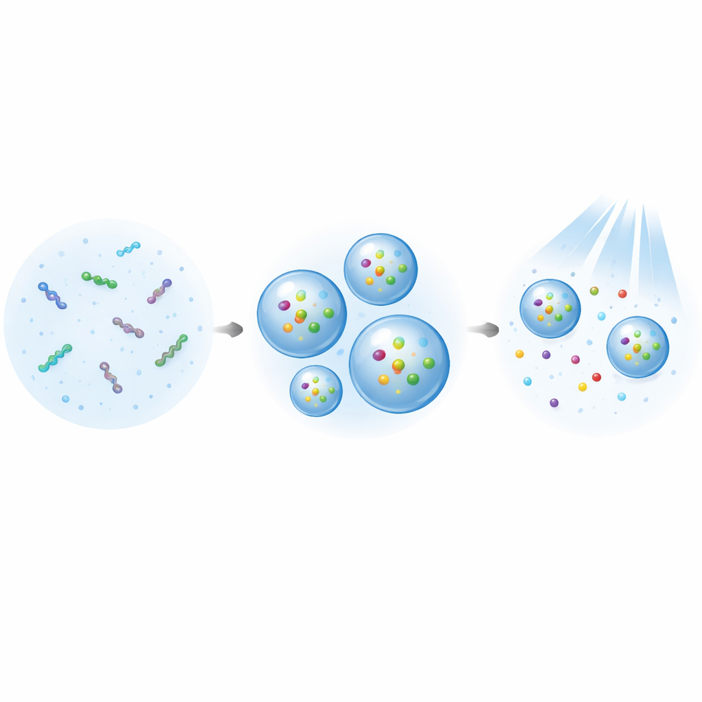
In this work, the team created “caged coacervating peptides” based on a histidine-rich protein from the hard, rubbery beak of the Humboldt squid. On their own, these peptides can form dense droplets, known as coacervates, in water. The researchers modified one amino acid in the sequence and attached a removable “cage” made from a coumarin-based chemical group. While the cage is attached, the peptides readily gather into droplets under mild salt and pH conditions similar to those in biological fluids. When the cage is removed by blue light, changes in charge and molecular stickiness weaken the tendency to form droplets, allowing them to partially disperse.
Testing the Droplets and Tuning Their Behavior
The scientists carefully checked how their caged peptides behaved in solution. They confirmed that the caged version formed microscopic liquid droplets, while the same peptide without the cage did not, except under more extreme conditions. The droplets disappeared when treated with a compound that disrupts weak hydrophobic contacts, a hallmark of true liquid phase separation. Using light, the team showed that the cage could be removed within seconds, and that this uncaging was tightly linked to the dose of light delivered. Initially, only about one-third of the droplet material dissolved when illuminated, suggesting that some interactions between the peptide chains and cage groups remained strong even after partial uncaging.
Making a Better Light-Dispersing Droplet
To improve light-driven breakup, the researchers introduced a second peptide design that weakened specific stacking interactions between aromatic side chains, making the droplets less tightly held together. This new peptide still formed droplets, but slightly less efficiently and with somewhat smaller particle sizes. Crucially, when exposed to blue light, these droplets fell apart much more readily: most of the peptide left the droplet phase and returned to the surrounding solution. This showed that carefully dialing down stickiness inside the droplet can make it more responsive to the light trigger without losing its ability to form in the first place.
Capturing and Releasing Cargo Molecules
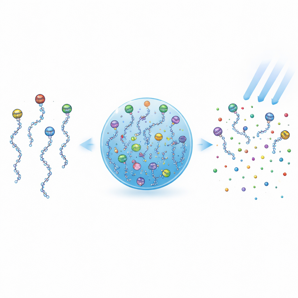
With a responsive droplet in hand, the team next asked whether it could store and release small cargo molecules on command. They chose a fluorescently tagged form of ATP, a common biological energy carrier, as a stand-in for potential drugs or signaling molecules. The improved peptide droplets took up about one-third of the ATP present in solution, concentrating it inside the droplet phase. When the sample was illuminated with blue light and then separated by centrifugation, most of the ATP was found back in the surrounding solution, showing that uncaging the peptides caused the droplets to release much of their cargo.
What This Means for Future Medicine and Research
In simple terms, the authors have built tiny, soft “containers” that can be filled with useful molecules and then opened partly by shining light. Because the trigger is blue light—a relatively gentle stimulus compared with harsh changes in temperature or acidity—this system may be kinder to living cells and delicate drugs. Although the droplets still need to be made more stable and precisely targeted, and only partial release was achieved, the approach points toward future drug delivery vehicles and light-switchable reaction chambers. By mimicking how cells naturally use liquid droplets to organize their chemistry, these designer peptides offer a versatile new tool for both basic biology and biomedical applications.
Citation: Bando, A., Kitamatsu, M., Kanazaki, Y. et al. Liquid–liquid phase separation by caged coacervating peptides. Sci Rep 16, 10464 (2026). https://doi.org/10.1038/s41598-026-40774-2
Keywords: phase separation droplets, light controlled peptides, drug delivery capsules, membraneless organelles, coacervate materials