Clear Sky Science · en
Dimeric magnetic dumbbell nanoparticles with selective immobilization of chromophores for improved tumor theranostics
Bringing Light and Nanotech Together Against Tumors
Cancer doctors increasingly rely on both seeing and treating tumors with light. Yet the very molecules that glow to reveal a tumor can interfere with those that generate toxic oxygen to kill it. This study introduces a tiny two-headed nanoparticle that carefully separates these light-responsive molecules in space, so they can team up for precise tumor imaging and photodynamic therapy without working against each other.
Why Light-Based Cancer Treatment Needs an Upgrade
Photodynamic therapy uses special drugs called photosensitizers that become deadly to cancer cells when illuminated with a specific color of light. When activated, they generate reactive oxygen species that damage tumor tissue while sparing most healthy cells. Many of these drugs also glow, which in principle should let doctors see exactly where the medicine accumulates and when to turn on the light. In practice, however, their glow is often weak, their signals overlap with tissue reflections, and the same absorbed energy must be split between making light and making toxic oxygen, limiting performance on both fronts.
The Energy-Leak Problem Between Glow and Therapy
To boost visibility, scientists often attach a bright fluorescent dye to the same platform as the therapeutic photosensitizer. But this introduces a hidden snag called energy transfer: if the two light-absorbing molecules sit too close and their colors overlap, one can silently siphon energy from the other. That can either dim the dye’s glow, making imaging difficult, or steal power from the photosensitizer, reducing its ability to kill cancer cells. Because most medical dyes and photosensitizers absorb and emit light in the same general visible range, it is nearly impossible to find a pair that completely avoids this unwanted energy exchange just by choosing different colors.
A Two-Headed Nanoparticle That Keeps Partners Apart
The researchers solved this by building a “dumbbell” nanoparticle made of two joined spheres: one of magnetite (iron oxide) and one of gold. Each half is coated and chemically tailored to host only one type of light-responsive cargo. The magnetite side is wrapped with small organic layers that selectively bind a near-infrared bacteriochlorin photosensitizer, optimized to generate reactive oxygen deep in tissue. The gold side binds a cyanine dye (Cy5) via strong sulfur–gold linkages, turning the same particle into a bright fluorescent beacon. Because the chromophores are anchored on different, physically separated surfaces, they are kept far enough apart to strongly reduce energy transfer, while still traveling together as a single nanoscale object. 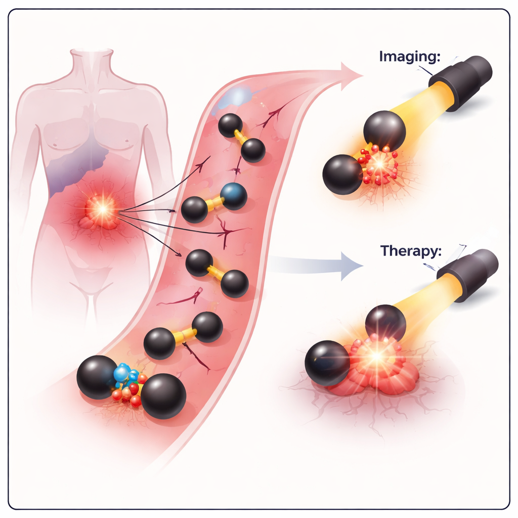
Stable Particles That Find and Enter Cancer Cells
The dumbbell design also addresses practical issues of drug delivery. The nanoparticles are small—under 30 nanometers in solution—and carry a hydrophilic polymer coating that helps them stay dispersed in blood-like fluids and evade rapid clearance by immune cells. Tests showed that these particles remain magnetically active, meaning they could also be steered or imaged by magnetic methods in future applications. In cell culture experiments with CT26 colon cancer cells, all three versions of the particles (carrying only the photosensitizer, only the dye, or both) entered cells efficiently and accumulated mainly in the cytoplasm and around the nucleus, rather than in the nucleus itself. Confocal microscopy revealed that, in the dual-loaded system, signals from the dye and the photosensitizer overlapped spatially, confirming that both cargos remained attached to the same nanoparticle inside the cells.
Switching On the Light: Safety and Tumor Cell Killing
The team next examined how safe and effective the system was. In the dark, even at relatively high concentrations, the nanoparticles showed little toxicity toward cancer cells, an essential requirement for any future therapy. When the cells were exposed to red and near-infrared light at clinically relevant doses, the particles bearing the photosensitizer produced strong, time-dependent cell death, consistent with robust reactive oxygen generation. Notably, the dual system with both dye and photosensitizer killed cancer cells more efficiently than the photosensitizer alone under the same conditions. This suggests that a small, controlled amount of energy transfer from the dye to the photosensitizer actually enhances treatment, without significantly undermining imaging. 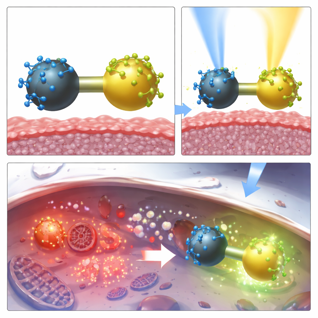
What This Means for Future Cancer Care
For a non-specialist, the take-home message is that the authors have engineered a tiny two-part particle that cleanly separates “see” and “treat” functions while still delivering them together to tumors. By physically spacing a glowing dye and a light-activated drug on opposite sides of a nanoscopic dumbbell, they largely avoid wasteful energy crosstalk, maintain brightness for imaging, and preserve or even boost the drug’s tumor-killing power. Because the particles are also magnetic, they could eventually support additional techniques like magnetic imaging or heat-based therapy. Overall, this work points toward smarter, multi-purpose cancer treatments where the same injected agent can help doctors locate tumors, monitor drug distribution, and then precisely destroy malignant cells with carefully timed light.
Citation: Chudosai, I., Ostroverkhov, P., Plotnikova, E. et al. Dimeric magnetic dumbbell nanoparticles with selective immobilization of chromophores for improved tumor theranostics. Sci Rep 16, 12101 (2026). https://doi.org/10.1038/s41598-026-40586-4
Keywords: photodynamic therapy, cancer nanomedicine, magnetite-gold nanoparticles, fluorescent imaging, theranostics