Clear Sky Science · en
Distinct functional networks derived from human induced pluripotent stem cell neuronal activity
Watching Brain Cells Learn to Talk
How do tiny collections of human brain cells learn to communicate, and why does their chatter sometimes fade with time? In this study, scientists grew networks of nerve cells made from human stem cells and listened in on their electrical activity for nearly two months. Their goal was to understand how simple, immature cells turn into organised networks that fire in unison, a process that underlies learning, memory, and many brain disorders.
From Skin-Like Cells to Miniature Brain Circuits
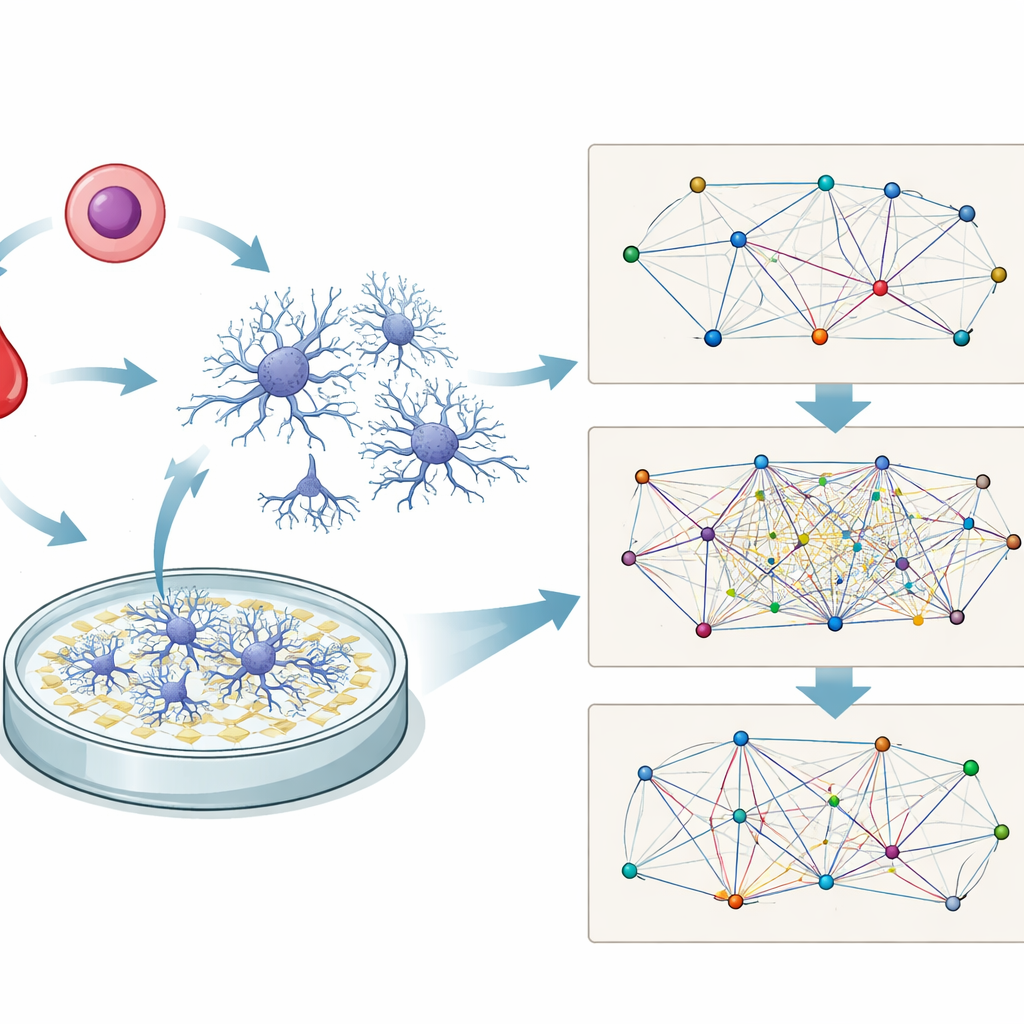
The researchers began with induced pluripotent stem cells, or iPSCs—adult human cells that have been reprogrammed to behave like embryonic cells. These iPSCs were coaxed into becoming excitatory brain cells and grown together with supportive star-shaped cells called astrocytes. Over days, the mixed cultures flattened into a thin layer and formed small clusters connected by long, branching processes that resemble the wiring of the brain. Using chemical stains that light up specific proteins, the team confirmed that these cells were forming both input and output sides of synapses, the junctions where nerve cells pass signals.
Following the Rise and Fall of Neural Conversations
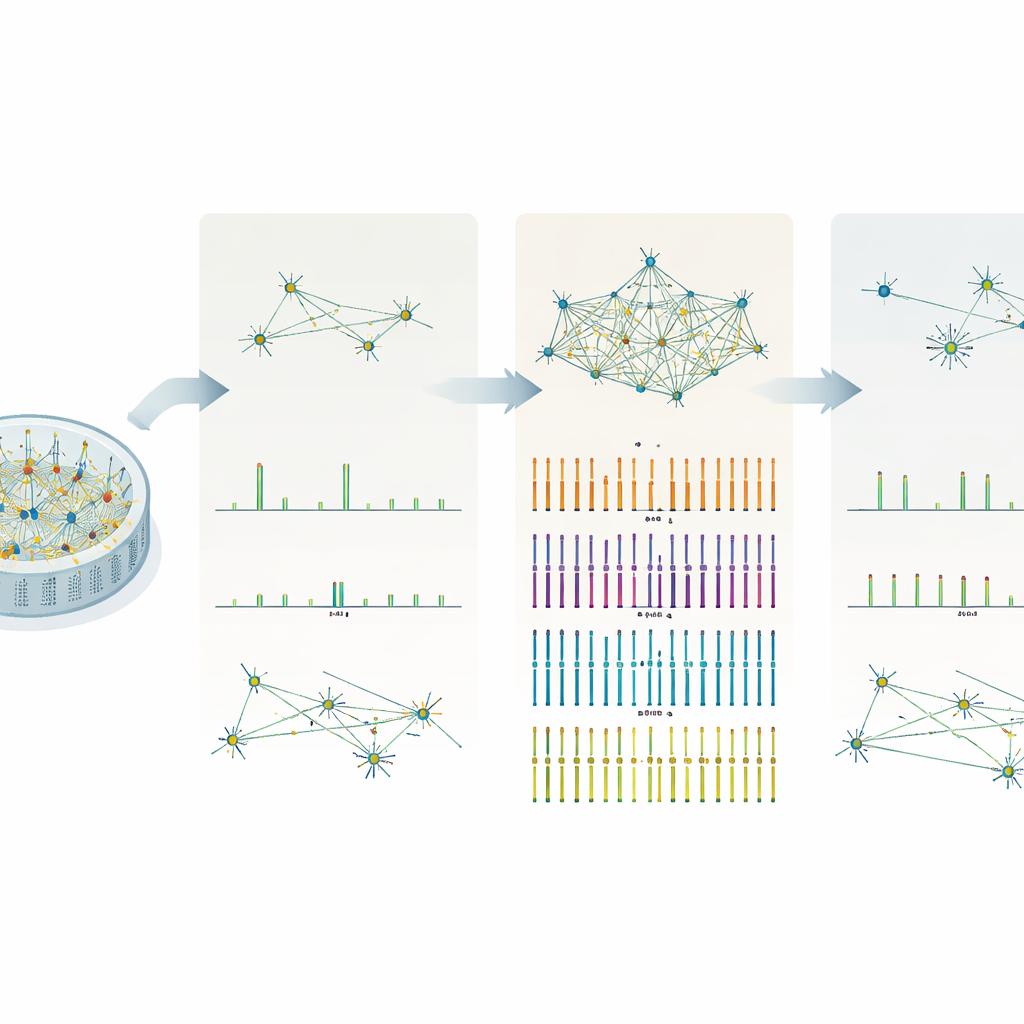
To track how these lab-grown brain cells behaved over time, the researchers used a multi-electrode array, a dish embedded with tiny sensors that can detect electrical spikes from many cells at once. They recorded five-minute snippets of activity from 24 such dishes on 21 different days, between day 18 and day 55 of culture. Early on, spikes were scattered and infrequent. Over the next week and a half, the total number of spikes, how often they occurred, and how often rapid “bursts” of spikes appeared all climbed steadily, peaking around days 24 to 28. After roughly a month in the dish, these measures began to decline, suggesting that the networks were losing some of their earlier coordinated drive.
Three Distinct Stages of Network Organization
Rather than looking only at raw spike counts, the team focused on how well different parts of the network fired together. They used a mathematical measure called phase locking to estimate how strongly activity at one electrode was linked to activity at others. This allowed them to build “functional connectivity” maps—abstract diagrams showing which regions tended to act in concert. When they grouped the data by age, three clear patterns emerged. In the earliest phase (days 18–23), most communication funneled through a single hub, and the network’s rhythm was weak and broadly spread. In the middle phase (days 24–28), connections became richer and more evenly shared across several hubs, and the network pulsed with a stronger, more regular rhythm. By the final phase (days 32–55), the maps simplified again, with fewer hubs and a weaker, less structured rhythm, indicating a partial breakdown or pruning of connections.
Linking Structure, Synapses, and Activity
The group also asked what was changing physically within the cultures while these electrical patterns evolved. Between day 21 and day 28, proteins that mark the presence of synapses—tiny contact points that allow neurons to talk—increased sharply. At the same time, markers of long, growing branches decreased, implying that the cells were shifting from building new extensions to refining and strengthening specific connections. A combined “maturation index” based on several synapse-related proteins more than doubled over this time. Together, these structural shifts matched the electrical data: as synapses multiplied and stabilised, network activity became more synchronous and organised before eventually tapering off.
Why These Mini-Networks Matter
For a general reader, the key message is that small, lab-grown networks of human brain cells go through recognisable life stages: they first spark to life, then become highly coordinated, and finally lose some of that order. This study shows that these changes can be tracked in detail using non-invasive electrical recordings and carefully chosen markers of cell structure. Because the stem cells can be made from any individual, such in-vitro networks offer a powerful way to study how genetic differences or potential treatments shape the wiring and timing of human brain activity, without needing to record directly from the brain itself.
Citation: Mehrkanoon, S., Rollo, B., Gu, J. et al. Distinct functional networks derived from human induced pluripotent stem cell neuronal activity. Sci Rep 16, 12659 (2026). https://doi.org/10.1038/s41598-026-40552-0
Keywords: induced pluripotent stem cells, neuronal networks, multi-electrode array, synaptic maturation, functional connectivity