Clear Sky Science · en
R3MV: a novel reliable system architecture for skin cancer classification using progressive heterogeneous multiblock model
Why this research matters for your skin
Skin cancer is among the most common cancers worldwide, and early detection can mean the difference between a simple procedure and a life‑threatening illness. Dermatologists increasingly turn to artificial intelligence (AI) to help spot dangerous moles in images taken with cameras and dermoscopes. But if different AI systems disagree on the same skin spot, which one should a doctor trust? This study introduces a new way to combine several AI viewpoints into a single, more reliable decision about whether a lesion is cancerous.
The problem with trusting a single smart machine
Most existing AI tools for skin cancer work by training one powerful model on one dataset. These systems can look impressive in lab tests, yet their performance often drops when they see images from new clinics, cameras, or patient groups. Prior evaluations in dermatology have highlighted weak external validation and poor generalization: models that work well on one carefully curated dataset may stumble in real-world hospitals. Because a wrong answer can delay treatment or cause unnecessary biopsies, the authors argue that relying on any single model is risky for clinical decision‑making.
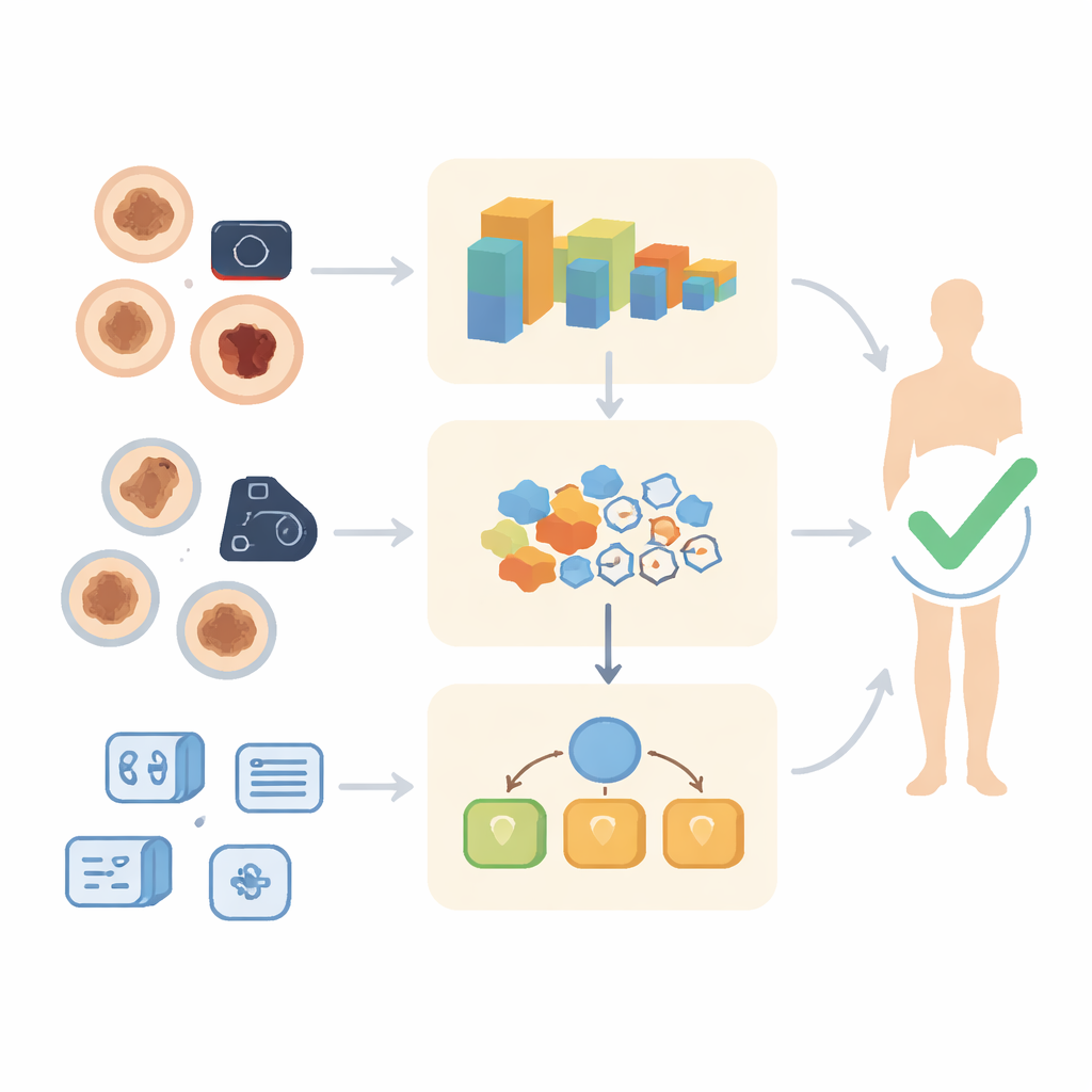
A new multi-voice voting system
To tackle this, the researchers propose a three-level architecture called R3MV, short for a reliable three‑level majority vote system. Instead of asking one AI model for an answer, R3MV runs the same skin image through three different decision paths. The first path uses a deep convolutional neural network that directly classifies the image. The second path feeds several different deep networks, merges the patterns they find, and passes these distilled features to a traditional classifier. The third path looks at the predictions made by multiple individual models and trains another classifier on these outputs. Finally, the system takes a majority vote across the three paths, so the final verdict reflects a consensus rather than a single opinion.
A smarter way to read skin images
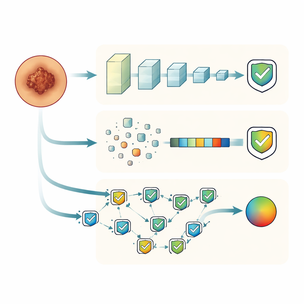
At the heart of the first path is a custom image model called the Progressive Heterogeneous Multi‑Block CNN (PHMBCNN). This network is built from several specialized blocks that each highlight different visual aspects of the lesion, such as fine textures or broader structures, in a progressive fashion from shallow to deep layers. The authors further enhance this model by adding a Gated Recurrent Unit (GRU), a type of memory module that helps the network capture relationships across channels and layers. This upgraded version, PHMBCNN‑GRU, serves both as a strong standalone classifier and as a critical feature source for the other paths in the system.
Letting multiple models learn together
The second path focuses on combining strengths from diverse image networks. The same lesion image is passed through PHMBCNN‑GRU and several popular pre‑trained models originally developed for general photographs. Each model extracts its own high‑dimensional “fingerprint” of the lesion. These fingerprints are concatenated and then compressed using techniques that remove redundant or unhelpful information, retaining only the most informative features. A multilayer perceptron, a classic layered neural network, then learns to classify lesions based on this refined joint description, effectively blending what each model “sees” into a single, sharper view.
Turning disagreements into a stronger answer
The third path, called decision‑level fusion, takes an even higher‑level view. Here, the authors treat each model’s predicted label as an input and teach a support vector machine to recognize patterns in the models’ agreements and disagreements. This meta‑classifier learns, for example, when certain model combinations usually indicate a benign spot or when a particular model tends to err on specific lesion types. Across all three paths, the researchers carefully preprocessed images—resizing, removing hair artifacts, and balancing classes—to better reflect the messy reality of clinical images captured by both dermoscopes and smartphones.
How well the system performs in practice
The team tested their approach on two widely used skin cancer image collections: HAM10000, which contains high‑quality dermoscopic images from clinical centers, and PAD_UFES_20, which includes smartphone photos with stronger artifacts such as dense hair and ink marks. The enhanced PHMBCNN‑GRU model alone improved accuracy compared with its baseline version on both datasets. More importantly, when all three paths were combined via majority vote in the R3MV system, performance rose further, reaching about 99% accuracy on HAM10000 and around 82% on PAD_UFES_20. Removing hair from lesions also consistently improved results, showing how crucial careful image cleaning is for automated diagnosis.
What this means for future skin checks
For non‑specialists, the key message is that this work moves AI for skin cancer from a single “black box” toward a panel of independent “opinions” that must agree before flagging a lesion as suspicious. While R3MV does not fully solve the challenge of making one system work equally well across all clinics and camera types, it shows that mixing different models and voting across them can yield more dependable answers than relying on just one. In time, such multi‑path systems could support dermatologists by offering more stable, explainable, and trustworthy assessments of which skin spots deserve closer attention or a biopsy.
Citation: Reshma, S.K., Reeja, S.R. R3MV: a novel reliable system architecture for skin cancer classification using progressive heterogeneous multiblock model. Sci Rep 16, 13599 (2026). https://doi.org/10.1038/s41598-026-40522-6
Keywords: skin cancer detection, dermatology AI, deep learning ensembles, medical image analysis, computer-aided diagnosis