Clear Sky Science · en
Application of treatment response assessment maps (TRAMs), based on delayed-contrast MRI for radiomic characterization of breast lesions
Why a New Breast Scan Matters
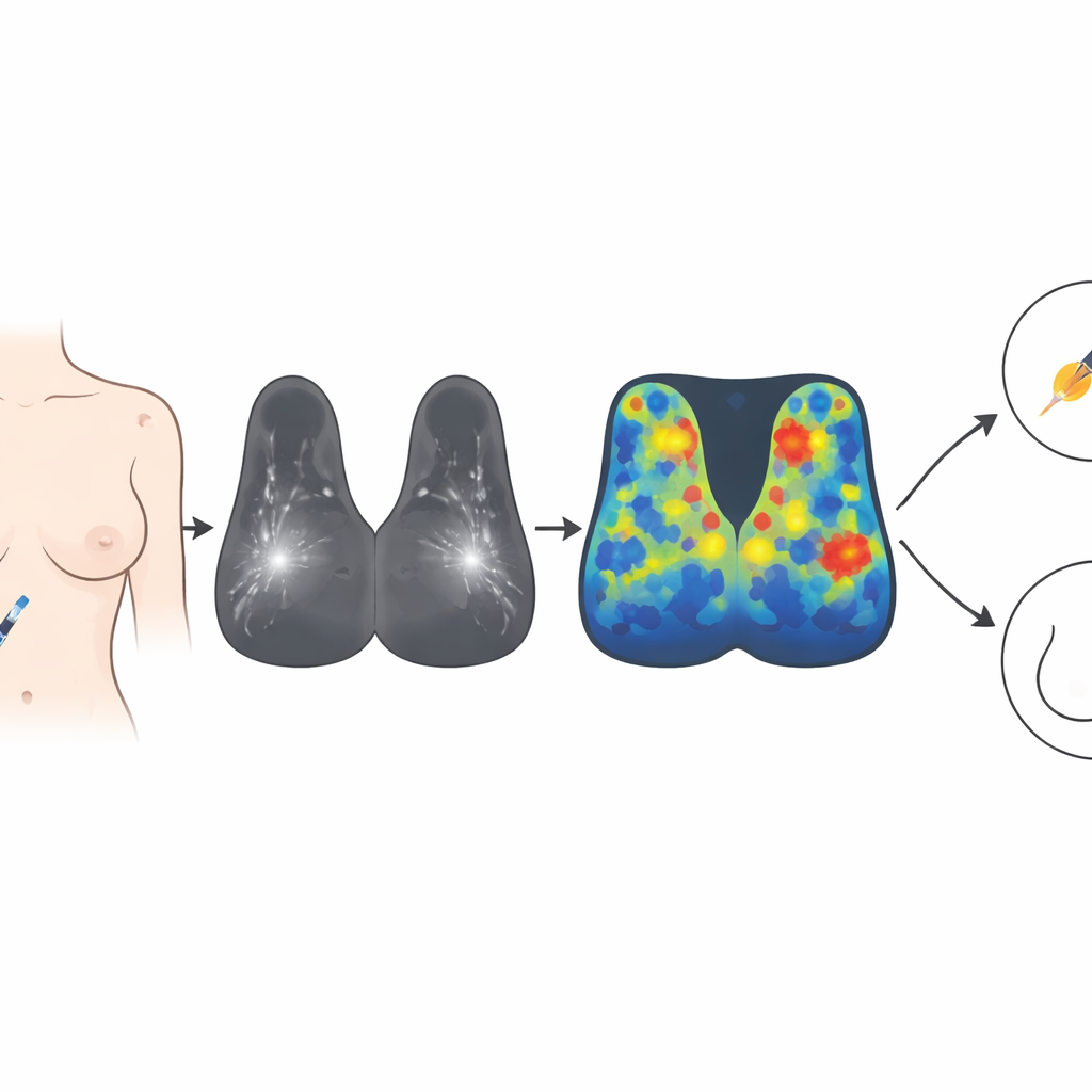
For many women, hearing that a breast MRI has found something "suspicious" leads straight to anxiety and, often, an unnecessary biopsy. While MRI is excellent at spotting possible cancers, it also flags many harmless changes in breast tissue. This study explores a new twist on MRI, called treatment response assessment maps (TRAMs), that may help doctors better tell dangerous tumors from benign findings, using information from how contrast dye slowly leaves the breast over time.
Seeing Beyond the First Few Minutes
Standard breast MRI relies on a contrast dye injected into the bloodstream and images taken over the first few minutes afterward. Cancerous tissue, with its abnormal blood vessels, often takes up and releases this dye differently from normal tissue. Radiologists look at how quickly areas in the breast brighten and then fade, but these patterns are not always clear-cut. Benign lesions can sometimes mimic cancer, leading to false alarms and extra tests. Radiomics, a data-driven approach that extracts dozens to hundreds of quantitative image features such as brightness patterns and shapes, has improved accuracy, but there is still room to reduce unnecessary biopsies.
Borrowing a Brain Technique for the Breast
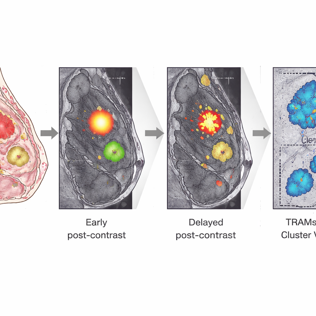
TRAMs were originally developed for people with brain tumors. Instead of focusing only on the first few minutes after contrast injection, TRAMs compare an early scan to a much later one, more than 20 minutes after the dye is given. By subtracting the two scans, TRAMs highlight areas where contrast has either built up or cleared out, revealing details about blood vessel function and the surrounding tissue environment. In brain cancer, this method can separate active tumor from treatment effects with very high accuracy. The authors of this study asked whether a similar delayed-contrast approach could help distinguish malignant from benign breast lesions.
How the New Maps Were Built
The researchers enrolled 135 women with 243 breast lesions that were either already known or suspected to be tumors. Each woman underwent a standard breast MRI, including a series of scans during the first few minutes after contrast injection, and then an extra scan taken about 22 minutes later while still in the scanner. Using advanced computer algorithms, the team carefully aligned the early and delayed images to correct for any movement, a crucial step to ensure that each tiny volume of tissue was compared accurately over time.
Turning Images into Numbers
From the regular contrast-enhanced images, the team extracted a large set of radiomic features describing how each lesion looked and how its brightness changed. From the TRAMs, they calculated simpler, physiologically focused measures: how much of the lesion showed delayed contrast clearance, how intense that clearance was, and how large the biggest connected region of clearing tissue was. They then used several machine learning methods to see how well these features could separate malignant from benign lesions, judging performance by sensitivity, specificity, accuracy, and the area under the ROC curve, a summary measure of diagnostic power.
What the New Maps Revealed
All malignant lesions showed a clear signature of contrast clearing on the TRAMs, while benign lesions tended to show persistent contrast build-up or mixed patterns. Models based on standard MRI radiomics alone achieved solid performance, with overall diagnostic scores in the range expected from the current literature. However, TRAMs-based models consistently reached slightly higher accuracy, with some combinations of TRAMs features achieving top scores. Strikingly, a model based on a single, easy-to-understand TRAMs feature — the volume of the largest cluster of clearing tissue within a lesion — performed nearly as well as, or better than, complex models built from many traditional radiomic features. This suggests that how much tissue in a lesion participates in delayed contrast washout carries powerful information about whether that lesion is cancerous.
What This Could Mean for Patients
For patients, the key message is that adding a single delayed MRI scan and using TRAMs to analyze contrast clearance could help doctors better judge which breast lesions truly require biopsy. The study shows that TRAMs provide information that standard imaging does not capture, while remaining relatively simple and interpretable. Although the approach requires extra scan time and was tested in a single center with a modest sample size, the findings suggest that, in carefully selected cases, delayed-contrast TRAMs might reduce unnecessary biopsies without missing important cancers. In the long run, this kind of smarter imaging could mean fewer invasive procedures, more confident diagnoses, and more tailored care in breast cancer screening and follow-up.
Citation: Daniels, D., Cohen, K., Last, D. et al. Application of treatment response assessment maps (TRAMs), based on delayed-contrast MRI for radiomic characterization of breast lesions. Sci Rep 16, 12170 (2026). https://doi.org/10.1038/s41598-026-40472-z
Keywords: breast MRI, radiomics, delayed contrast imaging, breast cancer diagnosis, TRAMs