Clear Sky Science · en
The performance of 68Ga-DOTA-IBA PET/CT for detecting bone metastases compared with 99mTc-MDP bone scintigraphy
Why finding cancer in bones matters
When cancers such as breast, prostate, or lung spread, they often travel to the skeleton. These hidden deposits in the spine, ribs, or pelvis can cause pain, fractures, and even paralysis, but they may be hard to spot early. This study asked a practical question that affects many patients: can a newer whole‑body scan called 68Ga‑DOTA‑IBA PET/CT reveal bone spread more clearly than the long‑used bone scan performed with 99mTc‑MDP? The answer helps doctors choose the right test to map where cancer has gone and how best to treat it.
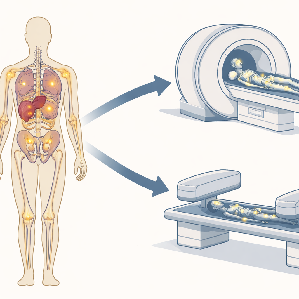
A closer look at two ways to scan bones
For decades, standard bone scans have relied on a small amount of radioactive tracer that homes in on active bone repair. A special camera then records the radiation to reveal suspicious “hot spots.” However, early metastases can hide inside the bone marrow, and many non‑cancer conditions—from arthritis to healing fractures—can also light up, making interpretation tricky. The newer approach, 68Ga‑DOTA‑IBA PET/CT, combines a different bone‑seeking tracer with a highly sensitive positron emission tomography scanner and a CT scan in one session. This promises sharper images and a better separation of true cancer deposits from everyday wear and tear in the skeleton.
Who was studied and how
The researchers reviewed records from 133 people with various cancers, including breast, prostate, lung, liver, and colorectal tumors, all suspected of having bone spread. Every patient underwent both tests—68Ga‑DOTA‑IBA PET/CT and 99mTc‑MDP bone scintigraphy—within one week, sometimes with targeted 3‑D SPECT images added to the bone scan. Experienced nuclear medicine physicians, unaware of each other’s readings, examined all the images. Using follow‑up scans and other imaging (such as CT or MRI), they decided which spots truly represented metastases and which were benign findings like bone islands or small cysts.
How well the new scan found bone spread
Across all patients, 1,453 bone metastases were confirmed. The PET/CT method detected nearly all of them: 1,423 lesions, or about 98 percent. In contrast, the bone scan with optional SPECT found 1,208 lesions, about 83 percent. This advantage held across cancer types and across different kinds of bone damage—whether the deposits appeared bone‑forming, bone‑destroying, or mixed on CT. At the level of individual patients, both tests identified almost everyone who had at least one metastasis, but PET/CT showed a richer map of how widely disease had spread, often revealing dozens of additional spots in the spine, ribs, pelvis, and skull that the bone scan missed.
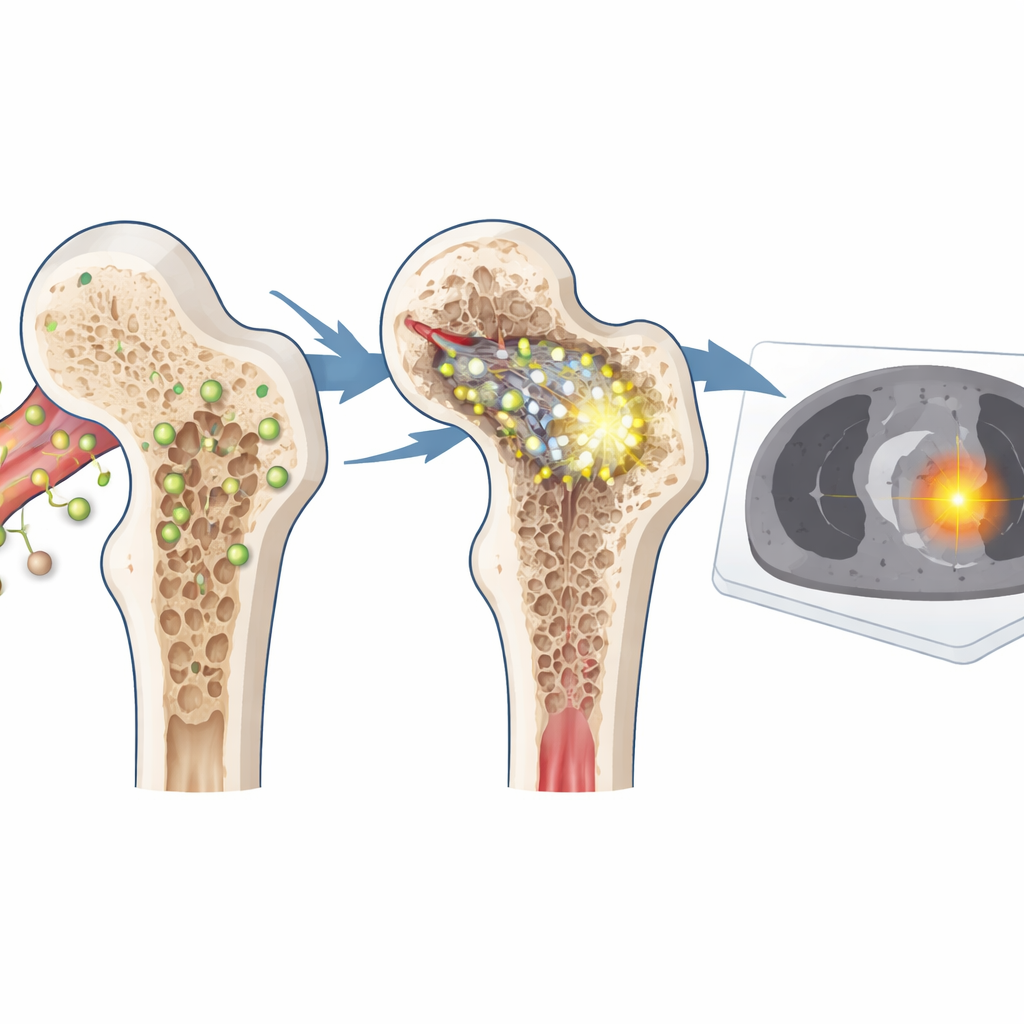
Separating cancer from harmless changes
Beyond simply seeing more lesions, the PET/CT method provided a numerical measure of how strongly each spot took up the tracer. In this study, true metastases picked up much more tracer than benign bone changes. When the researchers plotted these values, they found that a simple cut‑off level did an excellent job of telling malignant from non‑malignant areas. Using this threshold, the scan correctly distinguished most cancerous lesions from everyday bone quirks, offering a tool to clarify ambiguous findings that would otherwise worry patients and physicians.
What this means for patients
In plain terms, the study shows that 68Ga‑DOTA‑IBA PET/CT can draw a more detailed and reliable picture of cancer in the skeleton than the traditional bone scan. While both tests usually catch whether bone spread is present at all, PET/CT more accurately counts and localizes individual deposits and helps separate true metastases from look‑alike benign spots. That level of detail can refine staging, guide therapy choices, and support a “theranostic” strategy, in which the same type of compound can both image and treat bone disease. Although the work has limitations and needs confirmation in larger prospective trials, it suggests that for many patients facing the fear of cancer in their bones, this newer scan may offer clearer answers and better‑tailored care.
Citation: Lin, X., Zhang, N., Wang, R. et al. The performance of 68Ga-DOTA-IBA PET/CT for detecting bone metastases compared with 99mTc-MDP bone scintigraphy. Sci Rep 16, 10174 (2026). https://doi.org/10.1038/s41598-026-40411-y
Keywords: bone metastases, PET/CT imaging, nuclear medicine, cancer staging, bone scintigraphy