Clear Sky Science · en
An improved African vultures algorithm for multi-threshold optimization in chest X-ray image segmentation
Sharper views inside the chest
Chest X‑rays are one of the most common medical tests, but doctors still rely heavily on their eyes to separate healthy tissue from signs of disease. Computers can help by automatically dividing an image into meaningful regions, a process called segmentation. This paper introduces a new way to let a computer "slice" chest X‑ray images into several clean layers, so that areas such as lungs, bones, and abnormal shadows become easier to measure and track over time.
Why image slicing is harder than it looks
At first glance, splitting an image into dark and light parts seems simple: pick a cutoff brightness, and anything lighter than that belongs to one group, anything darker to another. Classic techniques, such as Otsu’s method and Kapur’s entropy, do exactly this and work well when only two groups are needed. Modern medical images, however, are far more complex. A chest X‑ray may need to be divided into several layers to separate lung tissue, ribs, heart shadow, and possible lesions. Trying to do this with many cutoffs quickly becomes a heavy calculation problem, and older methods slow down or fail to find the best way to partition the image.
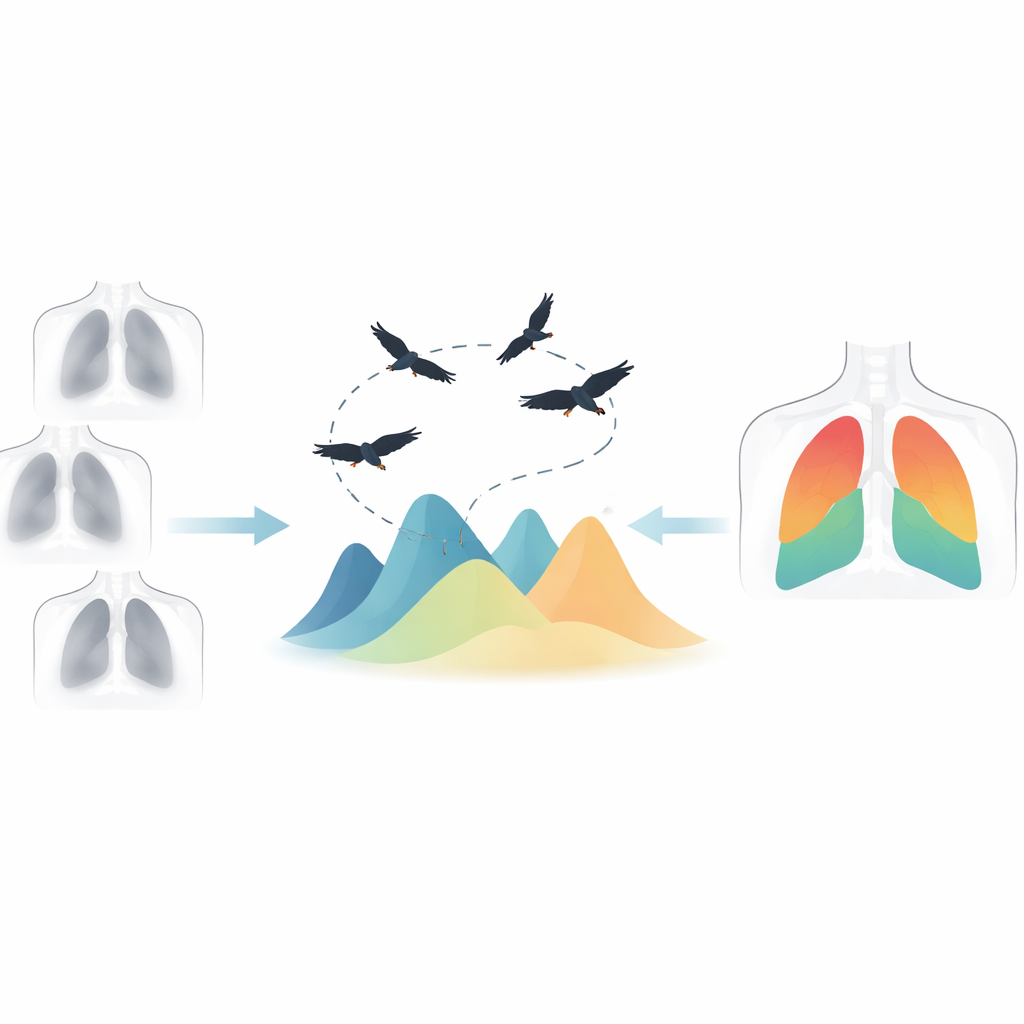
Borrowing strategy from scavenging birds
To tackle this challenge, the authors build on a recent idea from computer science inspired by African vultures. In nature, these birds roam widely when food is scarce and focus tightly when they close in on a carcass. The African Vultures Optimization Algorithm mimics this behavior: a group of virtual "vultures" explores a mathematical landscape searching for the highest peaks, which represent the best solutions to a problem. For image segmentation, each possible set of brightness cutoffs is one point in this landscape. The goal is to find sets that produce clear, informative slices of the X‑ray image.
Making the digital flock smarter
The original vulture algorithm can sometimes get stuck circling only one promising hill, missing better peaks elsewhere. The authors propose an Improved Multi‑Objective African Vultures Optimization Algorithm, or IMMOAVOA, to avoid this trap. First, they add an "average partial opposite" learning step: whenever the flock begins to cluster too tightly, new candidate solutions are generated by partially flipping their positions across the landscape and averaging them. This creates fresh, diverse options without discarding the progress already made. Second, they introduce an in‑depth exploration stage that refines especially promising candidates by nudging them in carefully controlled, shrinking steps, allowing the algorithm to home in on the best peaks instead of wandering indefinitely.
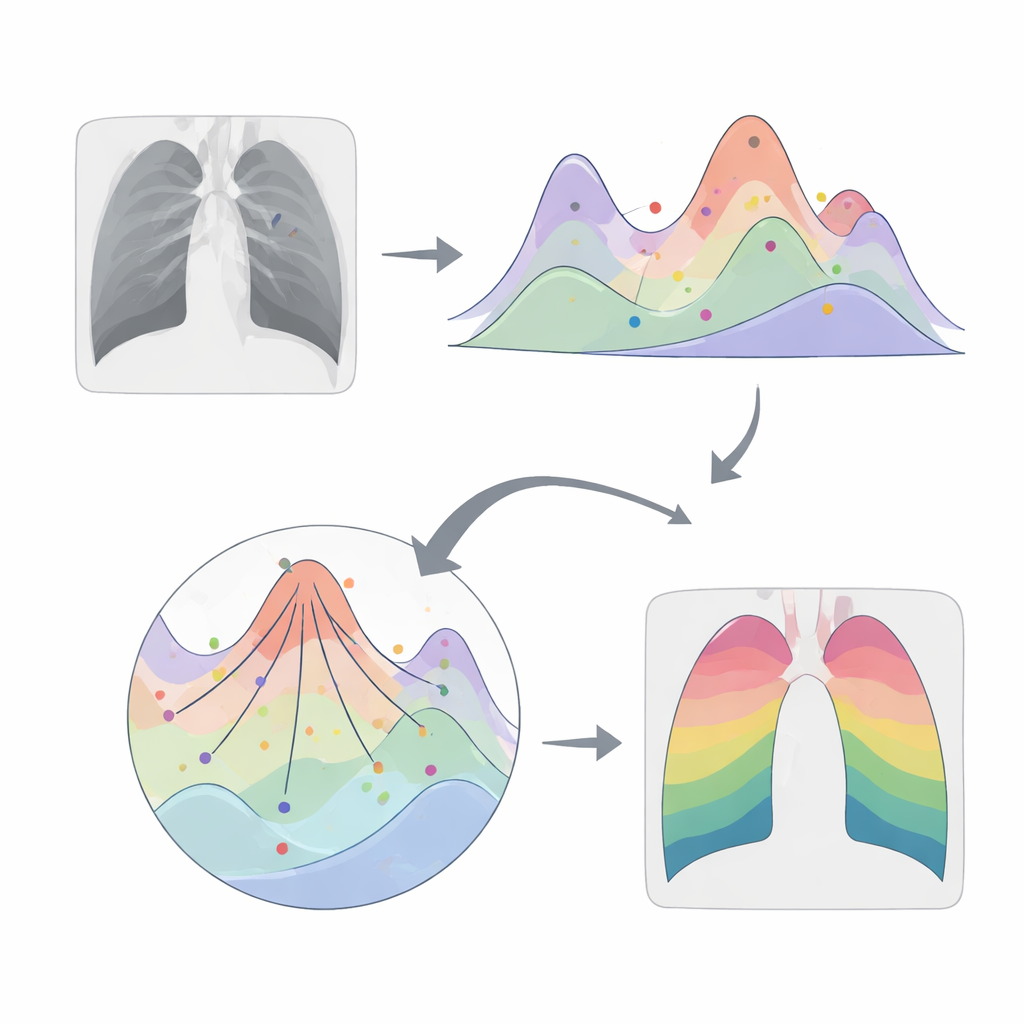
Balancing two ways of judging quality
Instead of trusting a single rule for what counts as a good segmentation, the new method combines two complementary viewpoints. One objective, related to Otsu’s method, rewards partitions that clearly separate bright and dark regions. The other, related to a two‑dimensional form of Kapur’s entropy, rewards partitions that capture as much information as possible from both the original brightness and its local neighborhood. IMMOAVOA searches for sets of thresholds that strike a good balance between these two goals, producing a collection of top‑ranked solutions rather than just one. The authors then apply these solutions to chest X‑rays from COVID‑19 patients and score the resulting images using several common quality measures that compare the segmented output to the original image.
How well the new approach performs
Before touching real X‑rays, the authors test IMMOAVOA on a standard suite of mathematical benchmarks used to judge search algorithms. Across measures that reflect both accuracy and diversity of solutions, the improved vulture method outperforms not only the basic version but also several well‑established multi‑objective algorithms. On actual chest X‑ray images, IMMOAVOA consistently yields clearer segmentations by three widely used indicators of image quality, while remaining competitive on a fourth measure that emphasizes overlap between regions. In practice, this means the method can carve chest X‑rays into multiple clean layers that preserve detail and structure, which is valuable for tasks such as outlining diseased lung areas or preparing data for further AI analysis.
What this means for medical imaging
For a non‑specialist, the takeaway is that the authors have designed a smarter and more flexible way for computers to "see" inside chest X‑rays. By coordinating a digital flock of search agents and judging each candidate solution from two different angles, their method can produce sharper, more informative slices of the image without an explosion in computing time. While the work is demonstrated on a relatively small set of chest X‑rays and still needs tuning before routine clinical use, it points toward more reliable, automated tools that could help radiologists detect and monitor lung diseases with greater precision.
Citation: Fu, Z., Liu, D., Gao, S. et al. An improved African vultures algorithm for multi-threshold optimization in chest X-ray image segmentation. Sci Rep 16, 10191 (2026). https://doi.org/10.1038/s41598-026-40174-6
Keywords: chest X-ray segmentation, medical image analysis, metaheuristic optimization, multi-thresholding, swarm intelligence