Clear Sky Science · en
Enhancing generative networks for chest anomaly localization through automatic registration-based unpaired-to-pseudo-paired training data translation
Why spotting lung problems early matters
Chest X-rays are one of the fastest and cheapest ways to look inside the chest, and millions are taken every day worldwide. Yet small or faint spots of disease can be hard to see, even for experts, especially when they blend into ribs, the heart, or other normal structures. This study explores how artificial intelligence (AI) can more reliably highlight suspicious regions on chest X-rays without needing painstaking, pixel-by-pixel hand-labeling by radiologists. That makes the approach especially appealing for hospitals and clinics with limited expert time and heavy patient loads.
The promise and trouble with today’s AI X-rays
Many existing AI systems for chest X-rays are good at answering a yes-or-no question such as “Is there pneumonia?” but much weaker at showing exactly where the disease is. One popular family of methods, called generative adversarial networks (GANs), learns to transform an image with disease into a version that looks normal. By subtracting the two, the algorithm can reveal suspicious areas. This is attractive because it does not require detailed markings of every lesion during training. However, the training data usually consist of separate sets of normal and abnormal images from different patients. Because body size, posture, and lung shape vary so much, the AI struggles to decide whether a difference between images is due to disease or simple anatomy. The result can be unstable performance and strange artifacts, such as ribs or normal tissue being distorted or erased.
Making virtual pairs from unpaired images
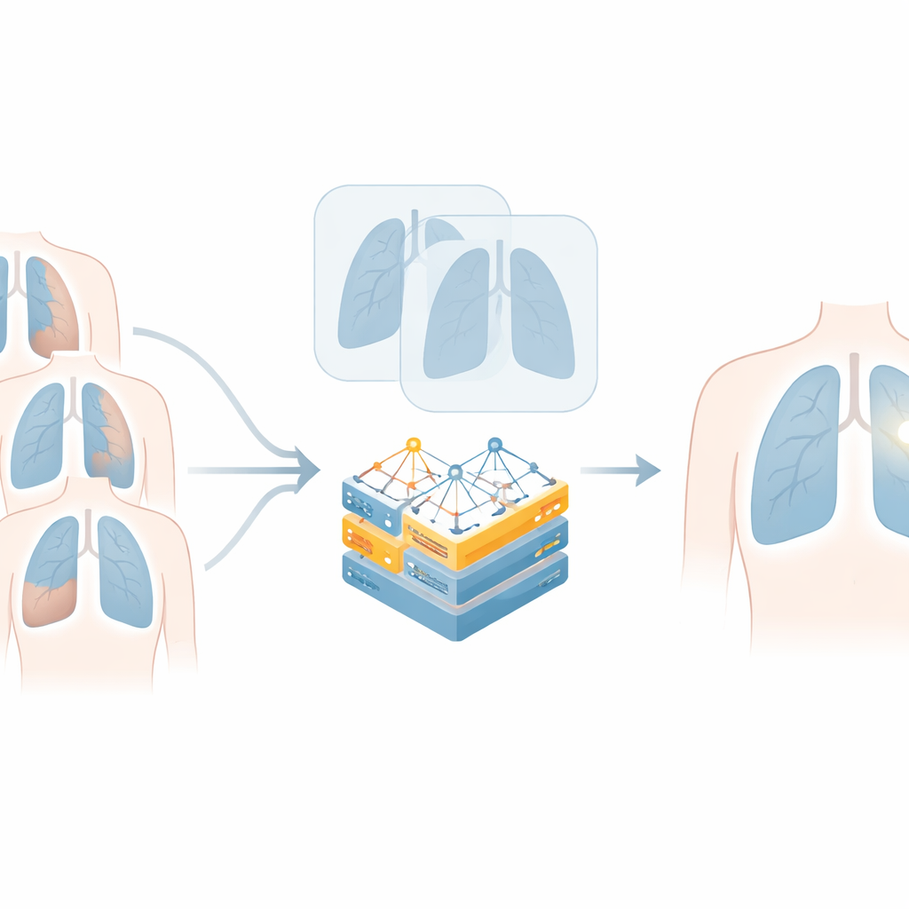
The authors tackle this key weakness by first forcing X-ray images into closer anatomical alignment before using them for learning and analysis. They introduce a two-step process, called IT-PR and its deep-learning extension IT-DPR, that effectively turns unpaired normal and abnormal X-rays into “pseudo-pairs.” In the first, training-free step, the method uses only the outlines of the lungs to map every point inside the lung to a common coordinate system. Each patient’s left and right lungs are segmented, stretched, and shifted so that equivalent internal locations line up across people, all relative to a single reference lung. This already reduces many gross mismatches in size and shape without relying on any disease information or neural network training.
Learning finer alignment with deep learning
Because simple geometric warping cannot perfectly match the subtleties of lung anatomy, the second step adds a learned, deformable registration. Here, a neural network is trained not on unrelated pairs of patients, but on pairs formed from the same patient: the original lung image and its pre-aligned version from the first step. In this way, the network learns only to correct coordinate differences, not to invent or remove disease. This refined registration produces smoother, more natural deformations and avoids two major failure modes seen in conventional registration methods: washing out real lesions when the “fixed” image is healthy, and creating unrealistic shading or kinks along lung borders when only crude masks are used.
Sharper AI spotting of tuberculosis and lung opacities
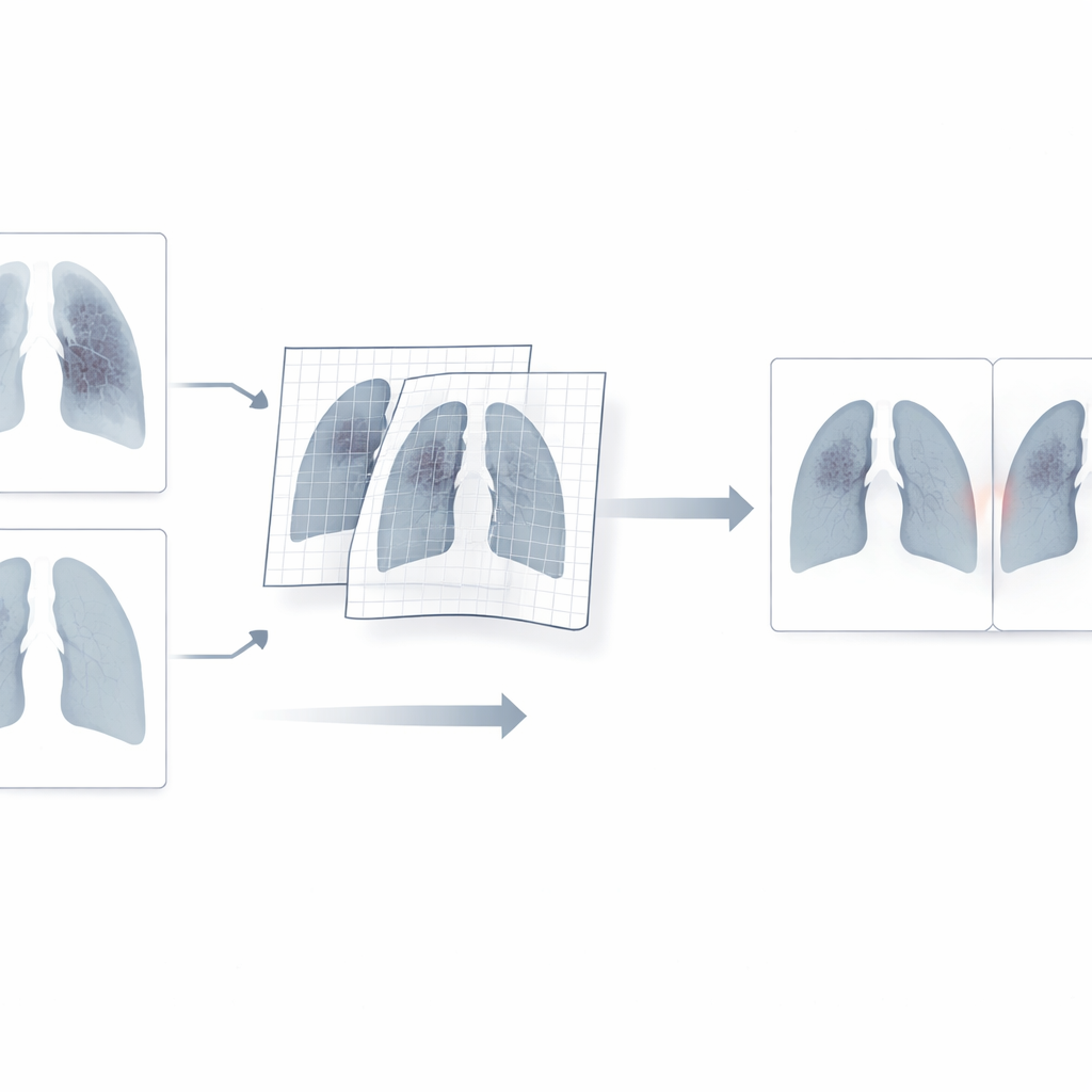
To test their approach, the researchers applied their registration-enhanced pipeline to two well-known chest X-ray problems: detecting tuberculosis and detecting consolidation, a type of lung opacity seen in pneumonia and other conditions. They plugged IT-PR and IT-DPR into two standard GAN-based translation models, CycleGAN and CUT, and compared them with the same models run in the usual way, as well as with a leading registration-aware generative model called Reg-GAN and popular registration tools such as VoxelMorph and SynthMorph. Using a patient-level “anomaly score” that summarizes how strongly an X-ray differs from its AI-generated normal counterpart, the new method greatly boosted performance. For tuberculosis, the best setup raised the area under the ROC curve from 0.755 to 0.928; for consolidation, it nudged an already high 0.964 up to 0.991. The approach also yielded more stable behavior across decision thresholds and noticeably cleaner, better localized highlight maps when compared with radiologist-provided bounding boxes.
What this means for patients and clinics
In practical terms, this work shows that carefully aligning chest X-rays before AI analysis can make a big difference in how reliably the system finds disease hot spots. By creating virtual normal–abnormal pairs without any pixel-level labels, the method keeps annotation costs low while improving both accuracy and interpretability. Although the added registration steps increase computing time to tens of seconds per image, this still fits within typical reading times for radiologists. The study’s framework is a prototype and depends on good lung segmentation, but it points to a powerful idea: when AI “looks” at medical images through a shared anatomical map, it becomes better at telling normal variation from true pathology, paving the way for more trustworthy, widely deployable tools for chest imaging.
Citation: Oh, S.J., Kim, K., Lim, C.Y. et al. Enhancing generative networks for chest anomaly localization through automatic registration-based unpaired-to-pseudo-paired training data translation. Sci Rep 16, 11125 (2026). https://doi.org/10.1038/s41598-026-39979-2
Keywords: chest X-ray AI, anomaly localization, medical image registration, generative adversarial networks, tuberculosis detection