Clear Sky Science · en
Real-time wavelet threshold denoising for laser speckle blood flow imaging
Clearer Views of Blood Flow
Doctors and researchers increasingly rely on cameras and lasers to watch how blood moves through tiny vessels in the brain, skin, and internal organs. One popular technique, laser speckle contrast imaging, can map blood flow over a wide area in real time, but its pictures are often grainy and unstable. This study introduces a faster, more reliable way to clean up those noisy images so that clinicians can see vessel details more clearly and trust the numbers that describe how blood is flowing.
How Laser Light Reveals Moving Blood
When a narrow beam of laser light hits living tissue, it scatters in many directions and forms a fine “speckle” pattern on a camera sensor. As red blood cells move, this speckle pattern subtly blurs and shifts over time. Laser speckle contrast imaging (LSCI) measures those tiny changes to estimate how fast blood is flowing in different parts of the field of view, producing a color-coded map of flow. Because it covers a large area in a single snapshot, LSCI is attractive for brain research, monitoring transplanted skin, and tracking microcirculation during surgery. Unfortunately, the same speckle effect that enables the method also creates strong noise that lowers image contrast and distorts the numerical link between the measured signal and the true speed of blood.
Why Existing Clean-Up Methods Fall Short
Researchers have tried several advanced image-processing tools to reduce speckle noise. Techniques such as non-local means and BM3D search for similar patches across an image and average them in clever ways, while variational mode decomposition breaks an image into several underlying components. These approaches can produce smooth backgrounds and good-looking pictures, but they come at a cost. They demand many tunable settings, are sensitive to how those settings are chosen, and require heavy computation. In practice, this means they often cannot keep up with high-resolution LSCI video streams and may fail when noise levels or imaging conditions change, limiting their usefulness in the operating room or at the bedside.
A Simpler Route to Stable Blood-Flow Maps
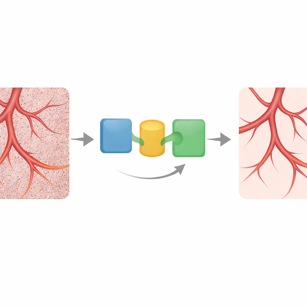
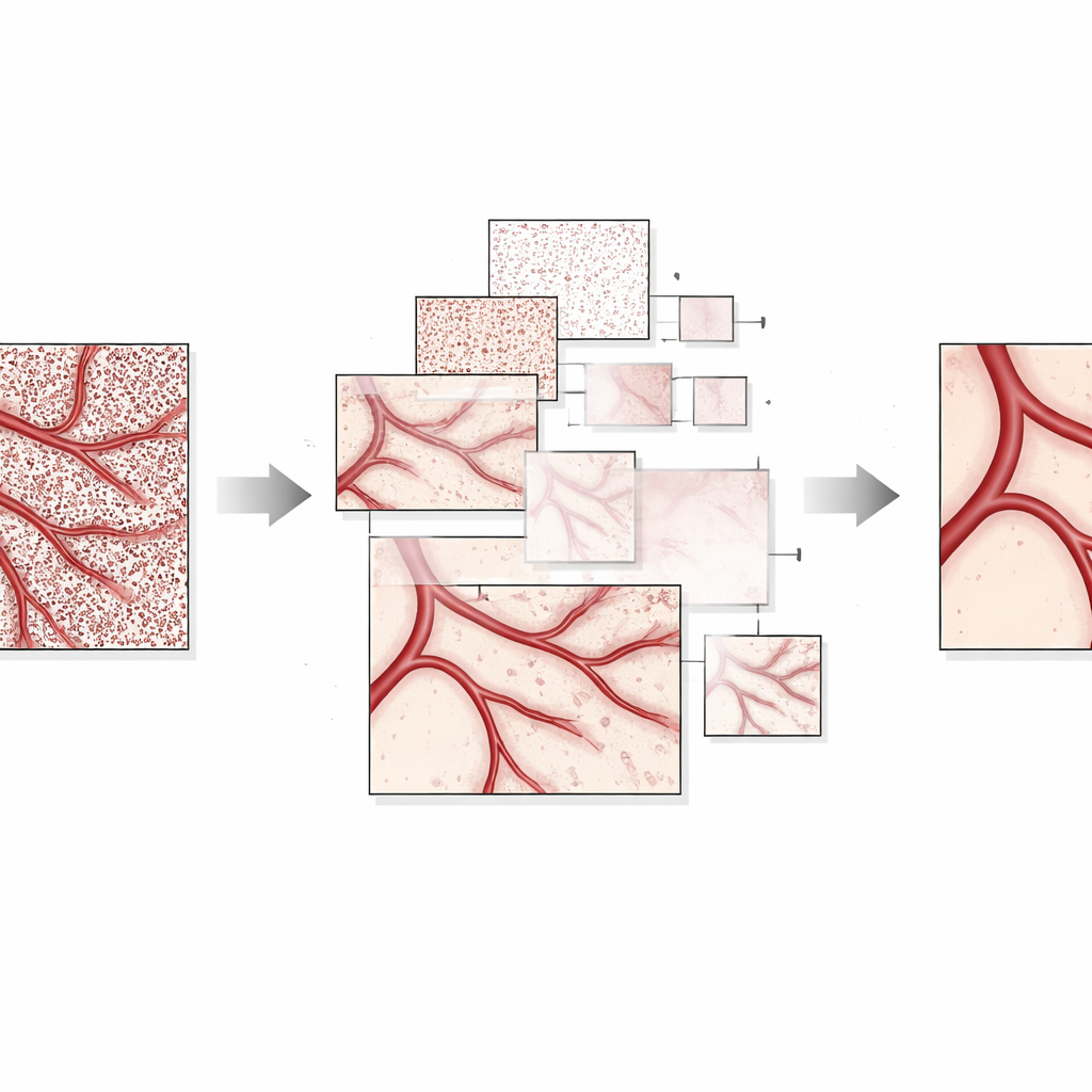
The authors propose a streamlined denoising method built around a common idea in signal processing: breaking data into different scales and gently shrinking the small, noise-like parts. First, they apply a mathematical trick, a logarithmic transform, that turns the speckle variations from a “multiplying” effect into an approximately “adding” one. This step makes the noise easier to handle. Next, they decompose the transformed image into several layers of detail using wavelets, which separate the broad vessel structures from fine-grained fluctuations. An adaptive rule, based on the Birgé–Massart principle, automatically chooses how strongly to shrink the high-frequency wavelet components associated with speckle while leaving the large-scale vessel patterns mostly intact. Finally, the image is transformed back and lightly stretched in brightness so that blood vessels stand out against the background.
Sharper Pictures, Truer Numbers, Real-Time Speed
To test their method, the team used both laboratory “phantoms” that mimic tissue and in vivo images from rabbit intestine. In the phantoms, where flow rates were precisely controlled, the new approach produced a nearly perfect straight-line relationship between the computed blood-flow index and the true flow speed, with the lowest error among all tested methods. In living tissue, the wavelet-based denoising delivered images that closely matched a high-quality reference made by averaging 100 frames, yet it did so using just three frames at a time. Objective measures of image quality improved, and the estimated blood-flow signals became smoother and more stable over time. Importantly, the algorithm processed full 4K frames in about 50 milliseconds on a graphics processor, far faster than BM3D and non-local means, and fast enough to support real-time visualization during medical procedures.
What This Means for Future Patient Care
By combining a simple set of fixed settings with automatic, image-driven thresholds, the proposed method reduces both speckle noise and the need for expert tuning. It preserves the fine branching pattern of tiny vessels while calming the random flicker that can mislead clinicians. Although some very subtle details may still be lost in scenes with large brightness swings, the balance of clarity, numerical reliability, and speed makes this approach a strong candidate for clinical laser speckle systems. In practical terms, it moves LSCI closer to being a routine tool for watching blood flow in real time, helping surgeons and physicians make faster, more confident decisions at the point of care.
Citation: Zhang, L., Yang, C., Liu, D. et al. Real-time wavelet threshold denoising for laser speckle blood flow imaging. Sci Rep 16, 10476 (2026). https://doi.org/10.1038/s41598-026-39846-0
Keywords: laser speckle imaging, blood flow mapping, medical image denoising, wavelet processing, real-time imaging