Clear Sky Science · en
Pneumonia detection from enhanced chest X-Ray images based on Double SGAN model
Why spotting pneumonia early matters
Pneumonia is a lung infection that can quickly become life-threatening, especially for children and older adults. Doctors often rely on chest X-ray images to see whether the lungs are clear or clouded by infection. But reading thousands of X-rays by eye is slow and subjective, and many clinics lack expert radiologists. This study explores how advanced computer vision can help: it describes a new way to train artificial intelligence so it can spot pneumonia on X-rays more accurately, even when there are far fewer images of healthy patients than sick ones.
The problem of uneven medical data
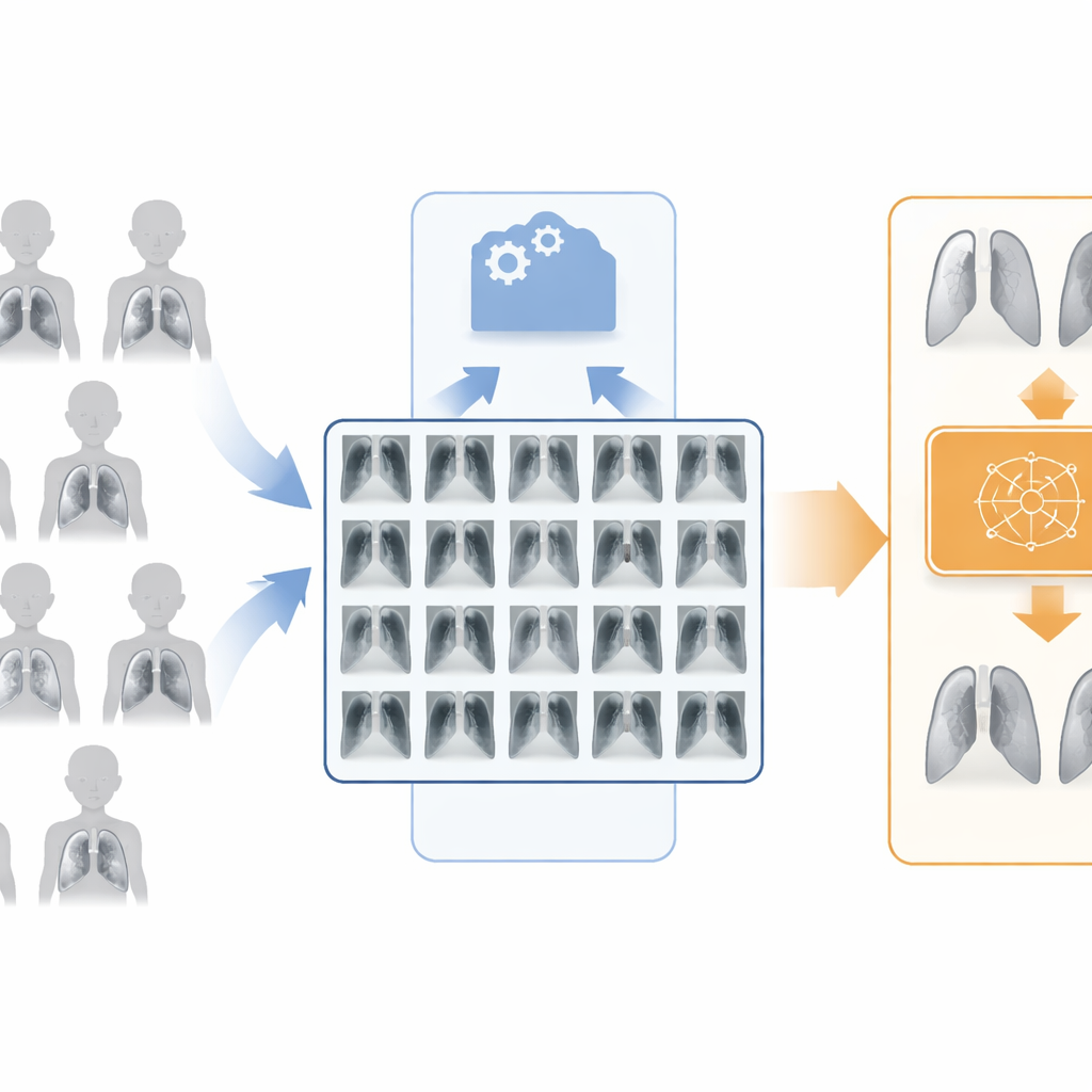
Modern image-recognition systems learn by seeing many labeled examples. In medicine, however, collecting and labeling images is difficult and time-consuming, and diseases are not evenly represented. In the pediatric chest X-ray collection used here, there are almost three times as many pneumonia images as normal ones. Standard deep-learning models trained on such imbalanced data tend to “play the odds”: they become very good at recognizing the common class and much less reliable for the rarer one. In practice, that can mean missing healthy children or, more dangerously, failing to flag sick ones. Traditional tricks like flipping or rotating images help only a little because they do not create truly new medical cases, just distorted copies of the same lungs.
Creating realistic extra X-rays on demand
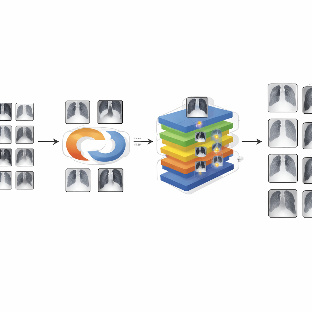
To overcome this, the authors turn to a family of models that can invent new images. They design an improved generative system called Double SGAN, which learns to produce highly realistic chest X-rays to bolster the minority class and balance the training set. One part of Double SGAN, the “generator,” starts from random noise and gradually shapes it into a synthetic X-ray, while another part, the “discriminator,” judges whether an image looks real or fake. The two components train together in a controlled rivalry. The authors reinforce this process with mathematical safeguards that keep the learning stable and prevent the model from collapsing into a few repetitive examples. They also add an internal “self-attention” mechanism so the generator can link distant regions of the image, helping it reproduce subtle lung patterns rather than mere blurry shadows.
Teaching the classifier where to look
Once Double SGAN has produced enough convincing normal images to balance the dataset, these synthetic scans are mixed with real ones to train a second model, called ResNet18-SA. This model is a streamlined image classifier built from many simple processing steps linked by “shortcut” paths, which help preserve details as information flows through the network. The key upgrade is a spatial attention module that learns to highlight the most informative regions of each X-ray—typically the lung fields—while downplaying background structures like ribs or the heart outline. By pooling information across the image and then reweighting it, the attention module nudges the network to concentrate on small, low-contrast patches that may signal pneumonia in a child’s lungs.
How well does the system work?
The researchers evaluate their approach on the publicly available Pneumonia-MNIST collection of 5,856 pediatric X-rays. They first check the quality of the synthetic images using established measures that compare the statistical “look” of generated and real images. Their improved Double SGAN sharply reduces the gap between fake and real scans, indicating that its X-rays are both diverse and lifelike. With these extra images in hand, they train ResNet18-SA and compare it against a range of well-known neural networks. On the original, imbalanced data, standard models reach accuracies in the low 90 percent range. After rebalancing the data with Double SGAN and adding spatial attention, ResNet18-SA achieves about 96 percent accuracy and similarly strong precision, recall, and F1 scores, surpassing heavier and more complex competitors while using relatively modest computational resources.
What this means for everyday care
For non-specialists, the core message is straightforward: by intelligently “imagining” extra, realistic X-ray images and then training a model that learns where to look inside each scan, the authors significantly improve automated pneumonia detection in children. Their system not only spots infected lungs with high reliability but also remains efficient enough for use in resource-limited clinics. Although the work is based on relatively low-resolution public data and focuses on a simple healthy-versus-pneumonia decision, it demonstrates how carefully designed generative models and attention mechanisms can help AI make better use of scarce medical images. With further refinement on higher-resolution scans and more detailed disease categories, this approach could become a valuable assistant for frontline healthcare workers worldwide.
Citation: Xu, Z., Zhang, H. Pneumonia detection from enhanced chest X-Ray images based on Double SGAN model. Sci Rep 16, 9922 (2026). https://doi.org/10.1038/s41598-026-39785-w
Keywords: pneumonia detection, chest X-ray AI, medical image augmentation, generative adversarial networks, deep learning radiology