Clear Sky Science · en
Lymphatic‑specific magnetic resonance lymphangiography biomarkers for grading lymphedema in animal models
Why swollen limbs matter
Many cancer survivors live with long‑lasting swelling of an arm or leg, known as lymphedema. This buildup of fluid can cause pain, heaviness, skin changes, and trouble moving, yet doctors still lack precise tools to see how badly the lymph system is damaged. The study behind this article tested a new type of MRI contrast agent in rats to see whether it can draw a clearer picture of lymph vessels, help grade how severe lymphedema is, and lay the groundwork for better diagnosis and treatment planning.
Swelling after cancer care
Lymphedema happens when the body’s drainage network for clear tissue fluid—the lymphatic system—is blocked or damaged, often after surgery or radiation for cancers such as breast, head and neck, or gynecologic tumors. Fluid then accumulates, stretching tissues and triggering inflammation and scarring. Although MRI scans and other imaging methods can show some of this anatomy, a major drawback has been the lack of a contrast agent that lights up lymph vessels alone. The agents most often used in MRI also brighten veins and other tissues, cluttering the images and limiting their usefulness for scoring how advanced lymphedema is.
A new way to light up the lymph system
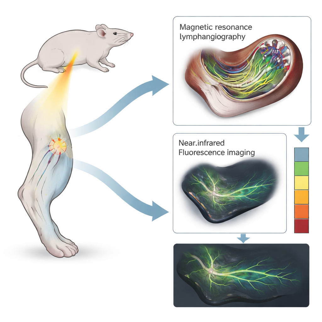
The research team worked with a specially designed iron‑oxide contrast agent called INV‑001. Unlike older iron‑based agents that mainly darken images, INV‑001 makes lymph vessels appear bright on certain MRI settings. The scientists created lymphedema in one hindlimb of rats by surgically removing key lymph nodes in the groin and behind the knee, then giving a focused dose of radiation to further disrupt drainage. After confirming that the affected ankle was at least one millimeter thicker than the healthy side, they injected INV‑001 into the skin between the toes and performed magnetic resonance lymphangiography (INV‑MRL). For comparison, they also imaged the same animals with a widely used optical method called near‑infrared fluorescence indocyanine green lymphography (NIRF‑ICGL), which traces the spread of a glowing dye just under the skin.
Seeing deeper and measuring more
Both imaging methods were able to show lymph vessels and changes caused by lymphedema, but they emphasized different features. The optical technique mainly revealed fine, superficial networks and striking patterns of “dermal backflow,” where dye leaked into the skin when vessels were overloaded or blocked. By contrast, INV‑MRL highlighted deeper, thicker lymph vessels and the extra “detour” channels that form as the system tries to bypass blockages, with almost no confusing bright signal from nearby veins. Experienced readers used visible patterns—such as straight, orderly vessels in mild disease and diffuse, hazy areas in more advanced cases—to assign lymphedema grades on a six‑level scale for each method. The grades from the two techniques matched extremely well overall, suggesting that INV‑MRL captures the same disease progression in a more anatomy‑rich way.
Turning images into numbers
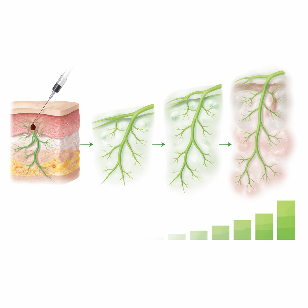
To move beyond expert impressions, the team also created a simple numerical marker called the threshold area ratio, or TAR. For each image, they drew a standard region covering the limb from ankle to hip and calculated what fraction of that area was bright enough to count as contrast‑filled tissue or vessel. As lymphedema became more severe, this fraction grew, reflecting both wider spread of contrast and more leakage into surrounding tissues. TAR values rose steadily with higher lymphedema grades for both INV‑MRL and NIRF‑ICGL and were strongly linked to each other. Importantly, in a small excretion study, INV‑001 had cleared from the injection site, lymph vessels, liver, and kidneys within 24 hours, suggesting that it does not linger in the body at the doses tested.
What this could mean for patients
In everyday terms, this work shows that a new iron‑based MRI contrast agent can selectively “paint” the lymph drainage network, reveal hidden deep‑lying vessels that other methods miss, and convert that picture into an objective score of how advanced lymphedema is. Because INV‑MRL avoids the overlapping signals and safety concerns associated with many gadolinium agents, it could eventually provide clearer guidance for surgeons planning lymph‑repair procedures and for clinicians tracking how patients respond to therapy. While these results come from animal models and will need to be confirmed in people, they point toward a future in which limb swelling after cancer treatment can be graded more reliably—and treated more precisely—than is possible today.
Citation: Cheon, H., Woo, DC., Chae, Y.J. et al. Lymphatic‑specific magnetic resonance lymphangiography biomarkers for grading lymphedema in animal models. Sci Rep 16, 10008 (2026). https://doi.org/10.1038/s41598-026-39610-4
Keywords: lymphedema, lymphatic imaging, MRI contrast agent, lymphatic vessels, disease grading