Clear Sky Science · en
A geometric function theoretic approach to MRI distortion correction using Miller Ross Poisson series
Sharper Scans Through Smart Math
Magnetic resonance imaging (MRI) is a mainstay of modern medicine, but the pictures it produces are not always as reliable as doctors would like. Subtle bends in the magnetic field and quirks in the hardware can warp brightness across an image, making some tissues look too dark or too bright. This paper shows how an advanced branch of complex analysis—geometric function theory—can be turned into a practical tool to correct such distortions, leading to clearer, more trustworthy scans.
Why MRI Pictures Can Be Misleading
In an ideal world, identical tissues in an MRI scan would have the same brightness everywhere in the image. In reality, the signal often fades or strengthens gradually from one side of the scan to the other, or changes in a nonlinear way. These effects arise from nonuniform magnetic fields, coil sensitivity variations, and hardware imperfections. Existing correction methods, such as polynomial fitting, homomorphic filtering, or bias field models, can help but are often heuristic: they depend heavily on experience, may overcorrect, and can behave unpredictably when noise or distortion is strong. This motivates methods that are not just empirical, but guaranteed by mathematical structure to behave in a controlled and stable way.
From Abstract Functions to Image Correction
The authors work with a special family of complex functions known as Sakaguchi-type functions, enriched using a construction called the Miller–Ross-type Poisson series. In simple terms, they build a toolbox of functions whose behavior is tightly constrained: their growth, bending, and distortion are all bounded and well understood. Within this toolbox, the team derives sharp limits on the function coefficients, as well as properties of their inverses and related quantities. These results belong to geometric function theory, a field that links algebraic formulas with the shapes they trace in the complex plane. Although these tools may seem abstract, the key idea is that any transformation built from such functions will be inherently well behaved: it will not fold over itself, explode in magnitude, or introduce wild oscillations.
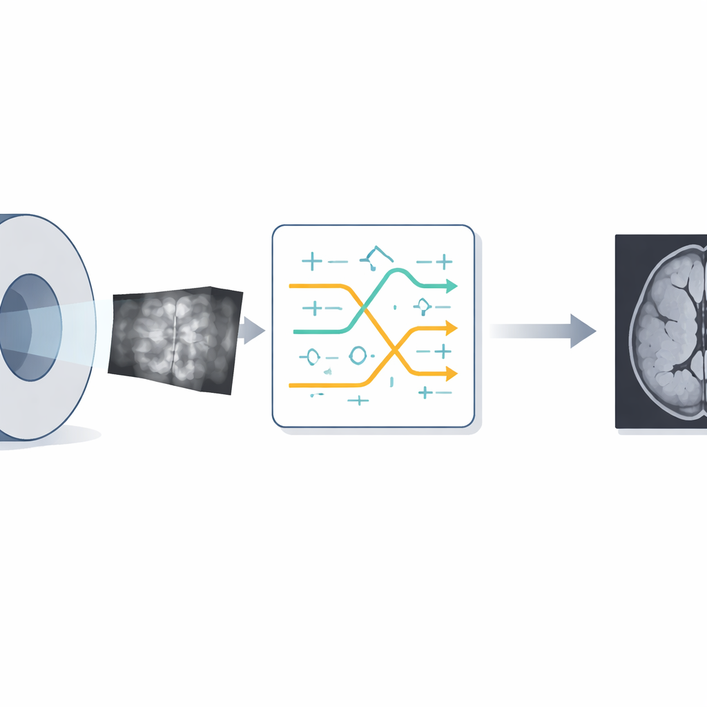
Designing a Safe Intensity Fix for MRI
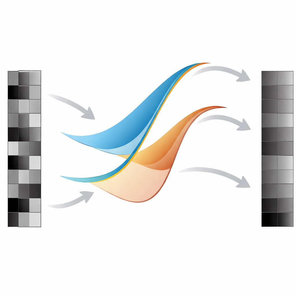
To turn this theory into an image-processing method, the authors model MRI intensity distortion as a nonlinear transformation of the true underlying brightness values. They then design a correction operator—a carefully chosen analytic function—that maps the distorted intensity back toward its original value. This operator takes the form of a low-degree polynomial whose coefficients must stay within the strict theoretical bounds derived earlier. By enforcing these bounds, the correction remains one-to-one and stable over the full intensity range, avoiding the overcorrection and structural damage that can happen with looser models. In practice, the team first simulates realistic distortions on normalized MRI images, then applies the analytic correction to each pixel’s intensity while preserving the overall image structure.
Putting the Method to the Test
The framework is evaluated on MRI data drawn from a public lung cancer collection (TCGA‑LUAD). The authors begin with reference images, apply a controlled nonlinear distortion to mimic real-world scanner imperfections, and then correct them using their analytic operator. They assess performance using standard image-quality measures: mean squared error (MSE), peak signal-to-noise ratio (PSNR), structural similarity (SSIM), and two no-reference perceptual scores, NIQE and BRISQUE. Compared with the distorted images, the corrected scans show lower error, higher PSNR, and improved structural similarity, indicating that fine anatomical details and tissue boundaries are better preserved. Even the perceptual metrics, which do not rely on a reference, show modest gains, suggesting that the corrected images look more natural as well as more accurate.
What This Means for Future Scans
In essence, the study demonstrates that carefully crafted analytic functions can serve as "safe" intensity correction tools for MRI, guided not by trial and error but by strict mathematical guarantees. By tying coefficient bounds and geometric properties directly to how pixel values are adjusted, the method reduces distortion while guarding against new artifacts. Although further clinical validation is needed, this work points toward a future in which advanced complex analysis supports more reliable medical imaging—and potentially other applications, from low-light photography to different types of medical scans—by ensuring that correction algorithms behave predictably and preserve the structures physicians need to see.
Citation: Manoj, S., Keerthi, B.S. A geometric function theoretic approach to MRI distortion correction using Miller Ross Poisson series. Sci Rep 16, 11639 (2026). https://doi.org/10.1038/s41598-026-39523-2
Keywords: MRI distortion correction, geometric function theory, analytic image enhancement, intensity inhomogeneity, medical imaging quality