Clear Sky Science · en
Longitudinal proteomic analysis of GCF in regenerative healing of molar furcation degree II defects treated with OFD, EMD, or A-PRF + : a pilot study
Why this matters for your gums and overall health
When dentists talk about “saving” a badly damaged tooth, they are often fighting disease hidden deep between a molar’s roots. This study looks inside that hidden space to see how different surgical treatments help the body rebuild lost support around teeth, and how a person’s general health—especially cholesterol and kidney function—can quietly steer the healing process. By tracking dozens of tiny proteins in the fluid that seeps from the gums, the researchers reveal early biological patterns that may one day help personalize gum surgery and improve the chances of keeping natural teeth.
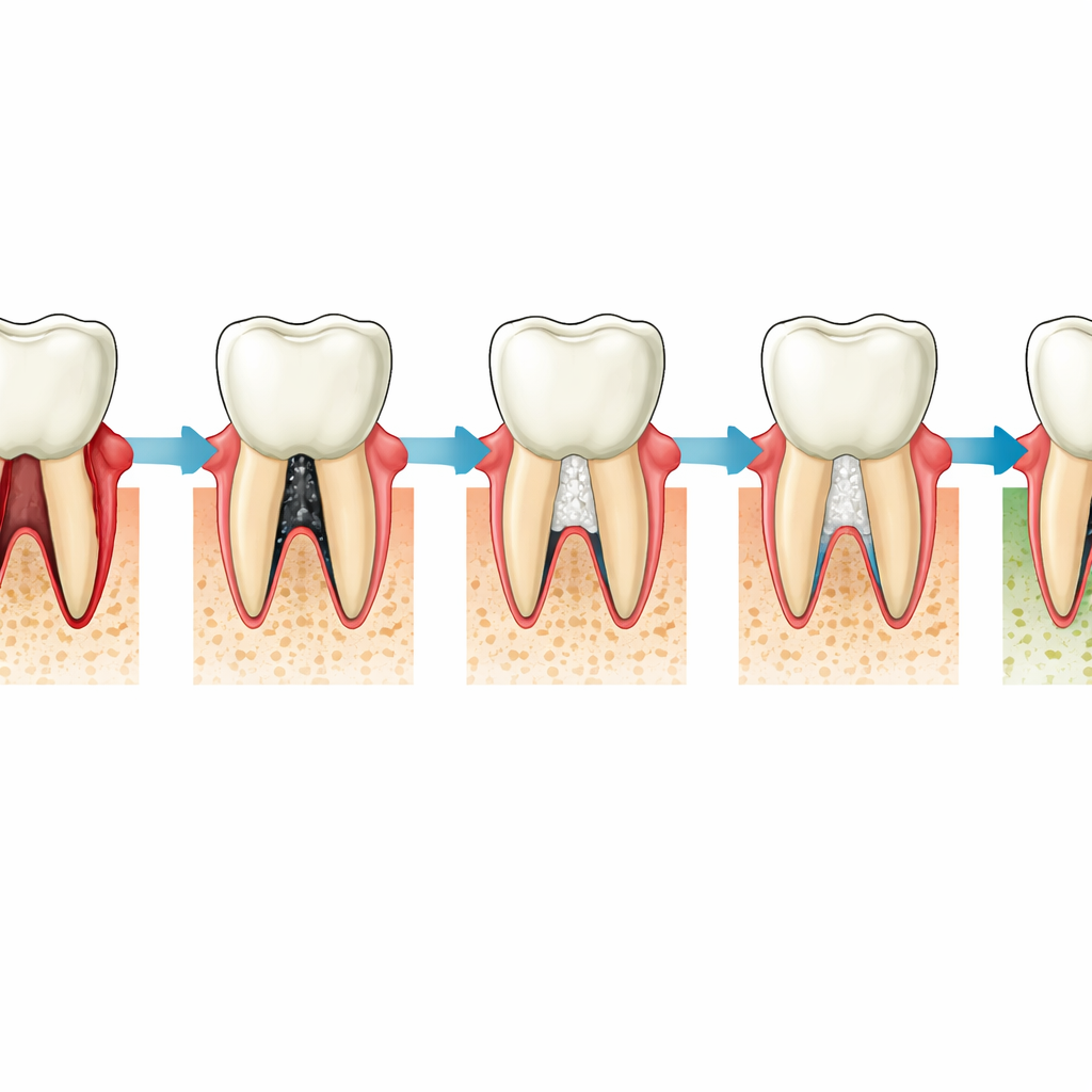
Deep trouble between the roots
Molar teeth can develop furcation defects, where bone is lost between the roots so that a “tunnel” forms under the crown. These problems are common in advanced gum disease and are notoriously hard to repair. The team studied degree II furcation defects, a stage where the damage is serious but the tooth can still potentially be saved with regenerative surgery. They compared three surgical approaches: simple cleaning and repositioning of the gum (open flap debridement, OFD), adding an enamel-based gel that encourages new attachment (EMD), and packing the defect with a sponge-like membrane made from the patient’s own blood clot (advanced platelet rich fibrin, A-PRF+). Seventeen patients were followed for six months after surgery.
Listening to healing through gum fluid
Instead of looking only at traditional measures like pocket depth and X‑rays, the researchers repeatedly sampled gingival crevicular fluid—the thin liquid that seeps from the space between tooth and gum. Using a highly sensitive laboratory panel designed to detect many proteins linked to inflammation, blood vessel growth, and tissue repair, they measured 46 different proteins at several time points from day 3 to month 6 after surgery. At the same visits, they scored how well the wound was closing using a modified early healing index. By matching protein levels with healing scores within each treatment group, they could see which biological signals rose or fell when healing went smoothly or poorly.
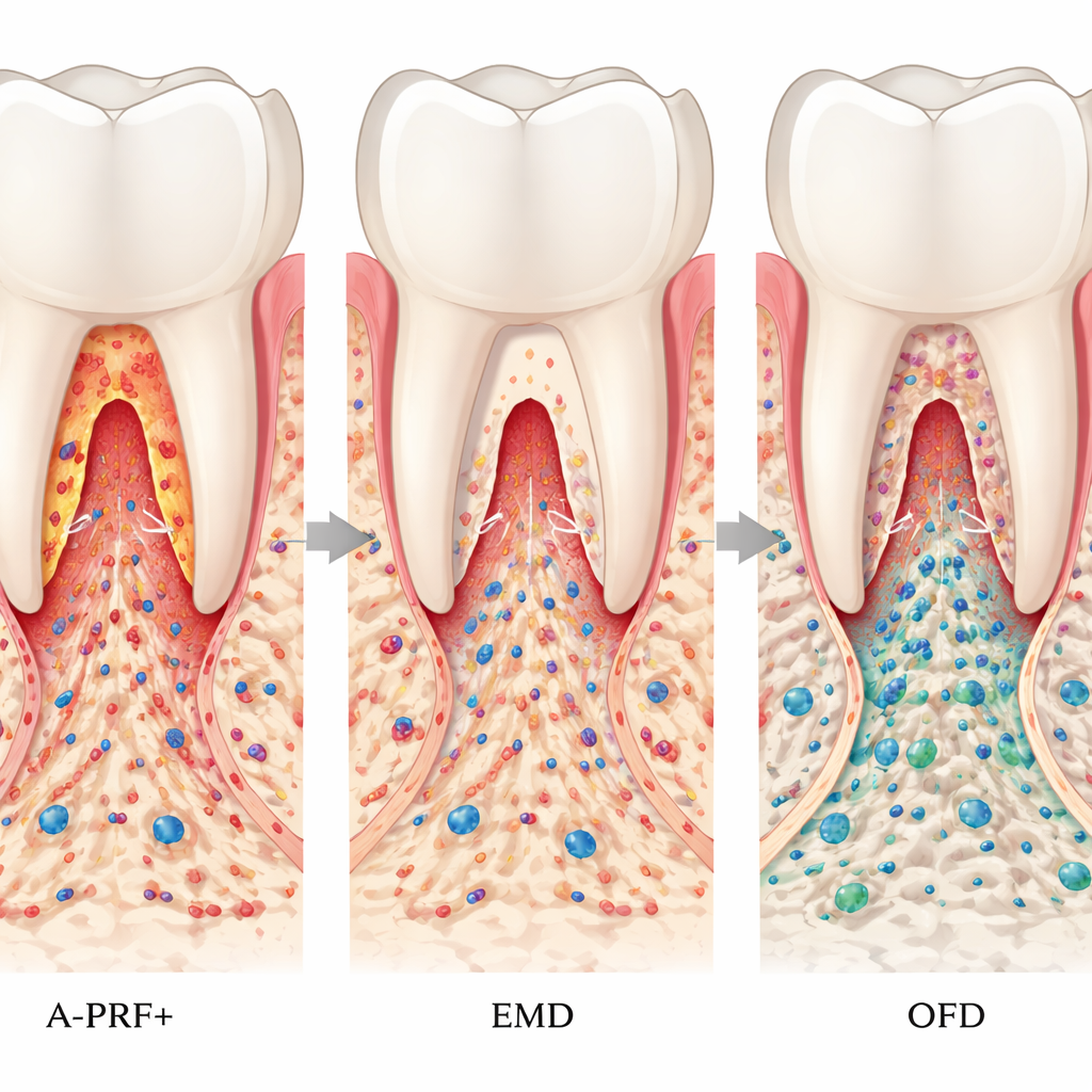
Different materials, different early healing stories
Clinically, the enamel-derived material (EMD) tended to give the quickest early wound closure, while sites treated with A-PRF+ closed more slowly and showed more variation between patients, even though several of these teeth later improved in furcation degree. In the earliest days after A-PRF+ treatment, proteins linked to cell death and acute inflammation—especially CASP‑8 and IL‑8—were higher when the wound looked worse. A week after surgery, A-PRF+ sites with slower healing still showed elevated CASP‑8 and a largely inactive form of a key growth factor (latent TGF‑β1), suggesting a drawn‑out inflammatory start before repair fully engaged. Later, around week 6 and month 3, other proteins involved in calming inflammation, shaping blood vessels, and remodeling tissue (such as ARG‑1, HGF, TRAIL, VEGFR‑2, TWEAK, LAP TGF‑β1, and CD40) showed distinct patterns between EMD, A‑PRF+, and OFD, indicating that each material steers healing along its own biological path even after the wound appears closed.
When blood chemistry slows gum repair
The team also asked whether a patient’s baseline blood values could forecast how well the tooth would recover. They looked at common markers measured in routine medical tests, including types of cholesterol and creatinine, a waste product that reflects kidney function. Higher levels of LDL (“bad”) cholesterol, higher total cholesterol carried in HDL, and higher creatinine at the start were all linked to less improvement in pocket depth, attachment, and bone level six months later, regardless of which surgical method was used. These findings support the idea that chronic metabolic and kidney problems do not just affect the heart or kidneys; they also subtly undermine the body’s ability to rebuild the fine structures that anchor teeth.
What this means for future gum surgery
Overall, the study shows that three common regenerative procedures for furcation defects can all lead to acceptable clinical results, but they do so with different early biological “signatures.” A-PRF+ appears to provoke a stronger and longer early inflammatory response yet may still support meaningful structural improvement in some difficult cases, while EMD favors quicker wound closure with protein patterns consistent with fast transition into repair and remodeling. Just as importantly, unfavorable blood cholesterol and creatinine levels are warning signs that healing may be limited. For patients, this points to a future where dentists may combine advanced gum surgery with personalized checks of blood and gum‑fluid markers to choose materials, protect fragile wounds, and coordinate care with physicians—improving the odds that even badly damaged molars can be kept for years to come.
Citation: Pitzurra, L., Stamatelou, E., Vasdravellis, D. et al. Longitudinal proteomic analysis of GCF in regenerative healing of molar furcation degree II defects treated with OFD, EMD, or A-PRF + : a pilot study. Sci Rep 16, 9832 (2026). https://doi.org/10.1038/s41598-026-39474-8
Keywords: periodontal regeneration, furcation defects, platelet rich fibrin, enamel matrix derivative, gingival crevicular fluid