Clear Sky Science · en
Associations between ventricular boundary shift integral and composite cognitive domains across the alzheimer’s disease continuum
Why brain scans matter for memory
As people live longer, many worry about whether forgetfulness is a normal part of aging or an early sign of Alzheimer’s disease. Doctors increasingly turn to brain scans to look for shrinkage, or atrophy, that might signal trouble ahead. This study explores a more dynamic way of reading those scans – called the Boundary Shift Integral, or BSI – to see whether it can better track changes in brain structure that go hand‑in‑hand with problems in memory, thinking, and language.
Looking across the path from normal aging to dementia
The researchers used data from a large international project that follows older adults over time. Participants fell into three groups: people with normal thinking, those with mild cognitive impairment (often a borderline stage between normal aging and dementia), and those already diagnosed with Alzheimer’s disease. Everyone took detailed tests of memory, executive skills such as planning and attention, and language abilities. They also had MRI brain scans at the start of the study and again 12 months later, allowing the team to see how their brains changed over that year.
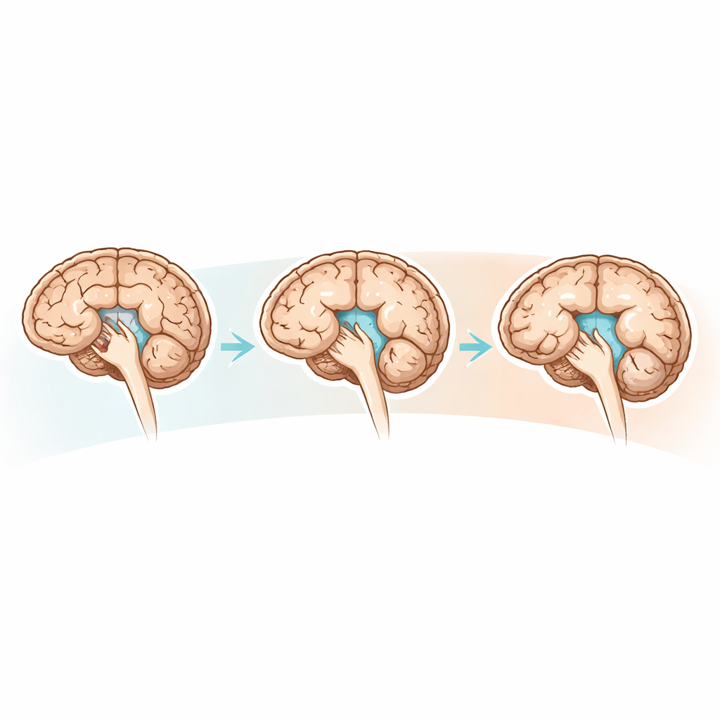
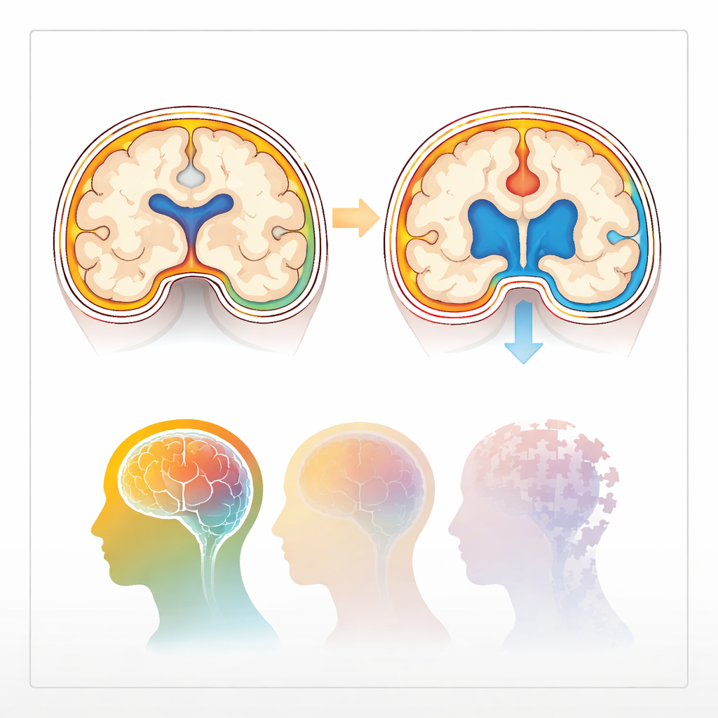
A moving picture of brain shrinkage
Traditional MRI measurements take a snapshot of brain volume at a single point in time – for example, how big the hippocampus (a key memory structure) is right now. BSI adds a time dimension: by carefully aligning two scans from the same person, it calculates how much the boundaries of the brain, its fluid‑filled spaces (ventricles), and the hippocampi have shifted. Expansion of the ventricles and thinning of brain tissue over 12 months provide a direct measure of how fast atrophy is progressing. The study compared these BSI measures for the whole brain, the ventricles, and the left and right hippocampus with standard volume measurements taken from the same scans.
Which brain changes track thinking skills best?
When the team linked brain changes to test scores, a clear pattern emerged. In people with mild cognitive impairment, BSI measures were strongly tied to performance in all three cognitive areas. Faster whole‑brain shrinkage and faster ventricle expansion were associated with worse memory, poorer executive function, and weaker language skills. Shrinkage of the hippocampus measured by BSI showed especially strong links to memory, echoing the central role of this structure in forming new memories. In people with established Alzheimer’s disease, BSI still detected meaningful relationships between brain atrophy and memory and thinking, particularly for whole‑brain and right‑side hippocampal changes, though the links were somewhat weaker, likely because many patients were already severely impaired.
How dynamic scans compare with standard measures
The researchers then asked whether BSI actually performs better than the usual static measures of brain volume. In people with normal cognition, neither approach showed clear ties between brain measurements and thinking skills, suggesting that at this stage changes are either too small or too subtle to pick up. In those with mild cognitive impairment, however, BSI consistently outperformed standard volumetrics in explaining differences in memory, language, and executive function. Standard volumes did show that larger brains and hippocampi, and smaller ventricles, were linked to better performance, but BSI captured these relationships more strongly and more reliably. In Alzheimer’s disease, whole‑brain volume and some hippocampal volumes still related to memory, yet again BSI tended to provide more consistent signals of how structural decline matched cognitive decline.
What this means for early detection and monitoring
For families and clinicians, these findings suggest that how quickly the brain changes over time carries more useful information than a single measurement of size. BSI turns pairs of routine MRI scans into a sensitive yardstick for tracking the spread of atrophy that underlies problems in memory, language, and higher‑order thinking, especially in the early, uncertain phase of mild cognitive impairment. While this method will not by itself diagnose Alzheimer’s disease, it could become an important part of a broader toolkit – alongside cognitive tests and other brain or fluid markers – to detect disease earlier, stage its severity more precisely, and monitor whether treatments are slowing the underlying damage.
Citation: Nasiri, H., Azimizonuzi, H., Khosravi, F. et al. Associations between ventricular boundary shift integral and composite cognitive domains across the alzheimer’s disease continuum. Sci Rep 16, 14092 (2026). https://doi.org/10.1038/s41598-026-39465-9
Keywords: Alzheimer’s disease, mild cognitive impairment, brain atrophy, MRI, cognitive decline