Clear Sky Science · en
Altered ciliary morphology reduces mechanosensation in a cystic kidney model as indicated by a mathematical model
How Tiny Hairs in the Kidney Help Protect Our Health
The cells lining our kidneys carry tiny hair‑like structures called primary cilia that sense the flow of urine. This study asks a deceptively simple question: what happens when those hairs change shape? By combining high‑resolution imaging of rat kidneys with a custom mathematical model, the researchers show that small structural changes in cilia can greatly weaken their ability to feel fluid flow, potentially helping cysts form in early cystic kidney disease. They also explore how something as basic as drinking more water might partially restore this lost mechanical signal.
Microscopic Hairs That Feel Flow
Primary cilia jut out from kidney tubule cells into the stream of passing urine. When urine flows, it bends these tiny hairs, and that bending is converted into internal chemical signals—especially calcium signals that help keep tubule size and function in check. In many cystic kidney conditions, genes linked to disease are active in these cilia, hinting that faulty flow sensing may be part of the problem. Yet it has been unclear exactly how changes in cilia shape inside real tissue alter the forces they feel. The authors used advanced three‑dimensional electron microscopy to visualize cilia in normal rats and in a cystic kidney model, and then built a mathematical model to translate those shapes into the drag forces produced by flowing urine.
When Straight Becomes Long and Curved
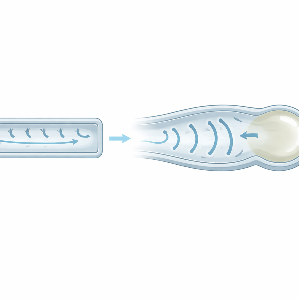
Imaging revealed that cilia in the cystic kidney model were not just a little different—they were markedly longer and more twisted than in healthy kidneys. In normal animals, cilia were relatively short and straight, like stiff antennae. In the cystic model, many cilia were extended and bent, resembling flexible hooks. Ultrastructural analysis showed why: in normal cilia, most of the internal skeleton consists of sturdy paired microtubules. In the diseased animals, over half of the ciliary length was made of single, thinner microtubules that are mechanically less rigid. This extended, softer “distal segment” made the cilia easier to bend and more curved, especially in tubules that were already dilated—early signs of cyst development.
A Mathematical Look at Invisible Forces
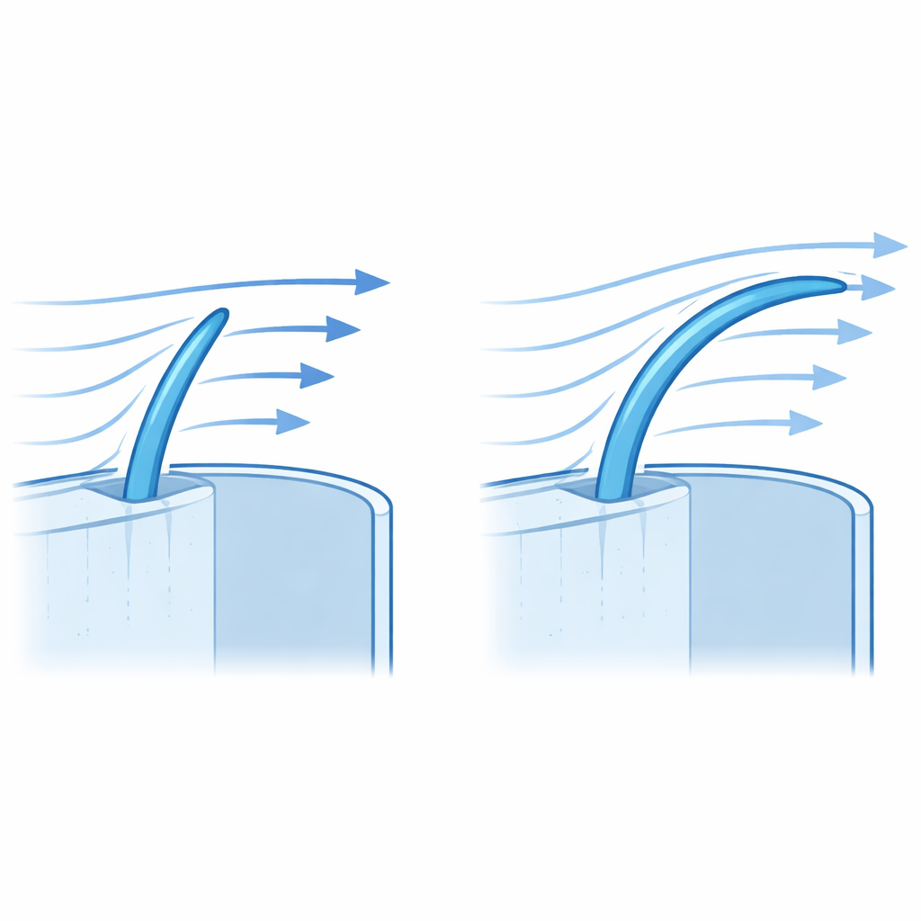
To understand what these shape changes mean for flow sensing, the researchers modeled how urine moves through a narrow tube and pushes on a cilium. They compared an idealized short, straight cilium to a long, quarter‑circle‑shaped bent cilium. Under gentle, smooth (laminar) flow, a straight cilium acts like a cantilevered rod: drag is highest near the tip, and bending strains concentrate toward the base where key signaling proteins, including the PC1/PC2 complex, reside. The model showed that when the same‑length cilium is instead long and bent, the effective surface facing the flow shrinks and the force is spread out along the curve. As a result, the total drag force on the curved cilium drops to roughly one quarter of that on the straight one, and the stress reaching the base—needed to trigger calcium entry—falls sharply.
How Much More Flow Is Enough?
The next question was practical: if bent cilia feel weaker forces, how much must urine flow increase to compensate? Using their equations and experimental data on how cylinders behave in slow flow, the authors estimate that cilia in the cystic model would need about 3.5 times higher flow to experience the same shear stress as straight cilia in a normal kidney. They then turned to an existing experiment in which cystic rats drank water sweetened with 5% glucose, a protocol known to drive animals to drink and urinate much more. In those high‑water‑intake animals, urine output rose several‑fold, above the model’s predicted threshold. In parallel, kidney tubules became less dilated, and cilia themselves became about 50% shorter and less curved—changes consistent with restored mechanical stimulation and healthier flow‑dependent control.
Why This Matters for Treating Cystic Kidneys
For a non‑specialist, the core message is that form and function are tightly linked even at the level of nanoscopic structures. When kidney cilia become too long, soft, and bent, they stop feeling the normal tug of flowing urine, so the cells lose an important feedback signal that helps prevent tubules from ballooning into cysts. The study’s mathematical model shows that this loss of sensation is not subtle: drag and shear can fall to a quarter of normal, unless urine flow is boosted several‑fold. High water intake can, in principle, supply that extra force, helping to shorten cilia and tame cyst growth—but only if the increase in flow is large enough, and always with attention to safety and comfort. More broadly, the work offers a quantitative framework to design therapies—whether through fluids, drugs, or other means—that restore the right mechanical cues to cilia, potentially slowing early cystic kidney disease.
Citation: Kumamoto, K., Kagami, H., Saitoh, S. et al. Altered ciliary morphology reduces mechanosensation in a cystic kidney model as indicated by a mathematical model. Sci Rep 16, 11485 (2026). https://doi.org/10.1038/s41598-026-39179-y
Keywords: primary cilia, cystic kidney disease, mechanosensation, urine flow, mathematical modeling