Clear Sky Science · en
Functional near-infrared spectroscopy identifies neural biomarkers of burnout in active-duty Police officers
Why brain clues to burnout matter
Burnout has become a familiar word, but for people working in high-stress jobs like policing, it can be a life-altering condition that affects judgment, health, and public safety. Yet today burnout is still diagnosed mostly through questionnaires and self-report, which makes it hard to spot early and to track objectively. This study explores whether subtle changes in brain blood flow, measured with a light-based headset, can reveal a reliable "neural fingerprint" of burnout in active-duty police officers—and whether computers can learn to distinguish officers at higher risk.
Stress on the job and the need for better tests
Burnout is more than just feeling tired. It rises from long-term workplace stress and shows up as deep exhaustion, a colder attitude toward one’s work, and a sense of reduced effectiveness. International agencies now recognize it as a major health concern, with huge economic costs. Police officers are a clear example of a vulnerable group: they face threats, trauma, public scrutiny, and disrupted sleep from rotating shifts. Studies link this kind of stress to heart disease, post-traumatic stress, higher suicide risk, and more frequent use of force. Despite this, assessments in the field mostly rely on written scales, which, while useful, can miss important biological changes and are open to personal bias.
Shining light on the working brain
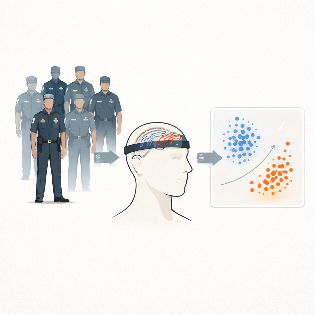
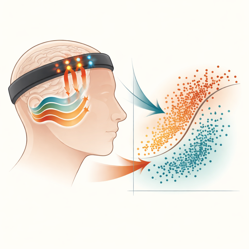
The researchers set out to create an objective burnout assessment tool starting with 33 active-duty officers in Taipei. Each officer filled out standard questionnaires on burnout, anxiety, and depression. The key burnout scale, adapted for Taiwanese workplaces, broke burnout into several types, including personal and work-related exhaustion. The team then fitted officers with a custom head-worn device that uses functional near-infrared spectroscopy (fNIRS). This technique sends harmless near-infrared light through the forehead and detects how much is absorbed by oxygen-rich and oxygen-poor blood, providing a window into activity in the prefrontal cortex, an area important for planning, self-control, and handling stress.
Putting thinking under pressure
While wearing the fNIRS headset, officers performed two kinds of demanding mental tasks. In a verbal fluency task, they had to quickly produce as many words as possible based on given sounds, a classic test of flexible thinking. In a mental arithmetic task, they solved a stream of math problems on a tablet. Each task included rest, active, and recovery periods so the team could watch how blood oxygen levels in specific frontal brain regions rose and fell with effort. From these signals, the researchers extracted dozens of features, such as how strongly blood oxygen increased during the task, how fast it changed at phase transitions, and how variable the signals were. These features were then fed into a type of machine-learning model called a support vector machine, which tried to learn the difference between officers with higher versus lower burnout scores.
Brain patterns that separate higher and lower risk
The most informative brain signals appeared during the verbal fluency task, especially from the right side of the prefrontal cortex. Two measures stood out: the size of the change in oxygen-rich blood and the change in oxygen-poor blood during the active phase. Officers in the higher work-related burnout group showed noticeably smaller shifts in both measures than their lower-burnout peers, suggesting reduced brain responsiveness or altered blood flow when under cognitive strain. Even though no single feature lined up neatly with questionnaire scores, combining just these two features allowed the computer model to distinguish higher- and lower-risk officers with about 91% accuracy in training data and 90% accuracy on a separate test set—far better than chance.
What this could mean for people at risk
To a lay reader, the key message is that burnout leaves detectable traces in how the brain supplies itself with oxygen when we think hard, and a simple wearable device can capture those traces in real time. By pairing light-based brain measurements with machine learning, this pilot study shows that it may be possible to build an objective screening tool that flags officers who are sliding into harmful levels of work-related burnout before problems fully surface. The authors caution that their sample was small and drawn from a single police district, so larger and more diverse studies are needed. Still, their results point toward a future in which burnout is tracked not only by how people say they feel, but also by how their brains quietly struggle to keep up under stress.
Citation: Chen, WY., Wang, WY., Huang, YH. et al. Functional near-infrared spectroscopy identifies neural biomarkers of burnout in active-duty Police officers. Sci Rep 16, 12477 (2026). https://doi.org/10.1038/s41598-026-38896-8
Keywords: burnout, police officers, brain imaging, near infrared spectroscopy, machine learning