Clear Sky Science · en
Ergothioneine promotes osteogenesis and angiogenesis through PI3K/AKT pathway and prevents glucocorticoid-induced osteonecrosis of the femoral head
Why protecting hip bones matters
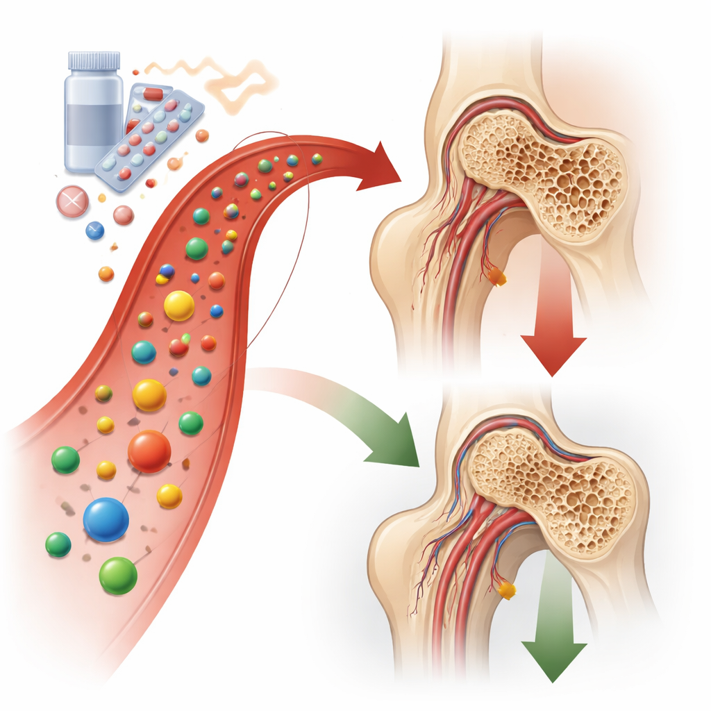
Many people rely on strong steroid medicines to control conditions such as autoimmune disease and severe inflammation. While lifesaving, long-term steroid use can quietly damage the hip joint, sometimes causing the round top of the thigh bone (the femoral head) to die and collapse. This painful condition, called osteonecrosis, often leaves joint replacement surgery as the only option, especially in younger adults. The study summarized here asks a hopeful question: could a naturally occurring dietary antioxidant, ergothioneine, help shield bone and blood vessels in the hip from steroid damage?
A hidden cost of powerful medicines
Osteonecrosis of the femoral head begins long before symptoms appear. High doses of glucocorticoids (a common class of steroids) disturb the cells that build bone and line blood vessels. They ramp up harmful oxygen byproducts inside cells, damage the tiny power plants called mitochondria, and trigger cell suicide. As bone-forming cells and blood vessels are lost, the femoral head weakens, blood flow diminishes, and the spherical bone can eventually cave in. Current treatments largely focus on pain relief or late-stage surgery; there are no widely used medicines that directly protect the bone and its blood supply from steroid injury.
A special antioxidant enters the scene
Ergothioneine is a sulfur-containing molecule found in foods such as mushrooms and produced by certain microbes. Our bodies actively transport and store it in organs that are vulnerable to oxidative stress, hinting at a protective role. Previous work linked ergothioneine to healthier brains and hearts, but its effects on bone were unknown. The researchers first used large-scale gene data from patients with steroid-related osteonecrosis to map molecular pathways involved in the disease and to match them with predicted targets of ergothioneine. This analysis highlighted a central growth and survival system inside cells, known as the PI3K/AKT pathway, as a potential bridge between the antioxidant and bone protection.
Testing bone and vessel cells under stress
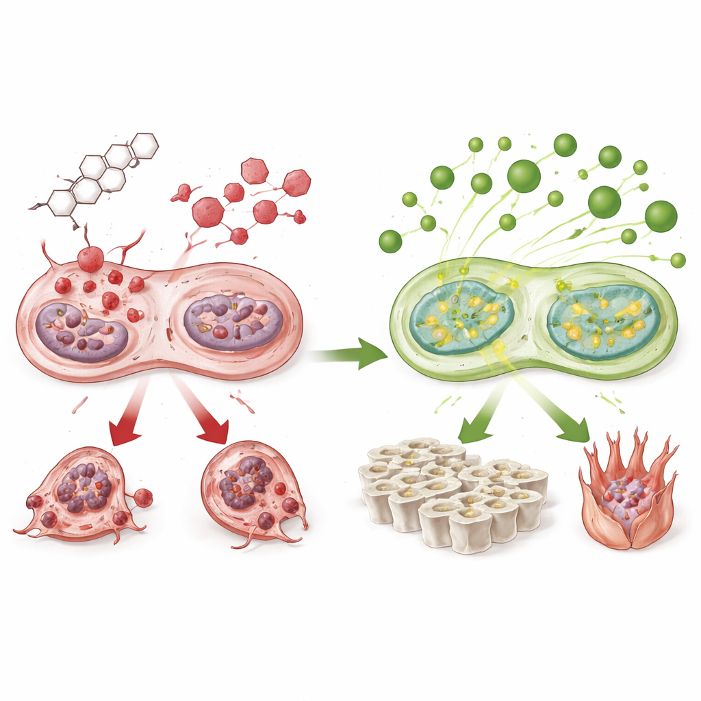
To probe this link, the team exposed two key cell types to a strong steroid in the lab: bone marrow stem cells that can become bone-forming cells, and endothelial cells that form blood vessel walls. Steroid treatment alone reduced these cells’ ability to mature into bone-making cells, lowered markers of bone formation, disrupted the formation of tiny vessel-like tubes, and increased signs of programmed cell death. Mitochondria became swollen and fragmented, and levels of damaging reactive oxygen species rose sharply. When ergothioneine was added before the steroid, many of these changes were reversed: bone and vessel cells survived better, maintained their ability to build bone and form tubes, produced less damaging oxygen, and showed healthier mitochondrial shape and energy signals.
Following the cell’s survival circuitry
Looking deeper, the researchers measured how strongly the PI3K/AKT pathway was switched on. Steroids dampened this pathway, reducing the activated forms of its main proteins. Ergothioneine restored their activation, coinciding with fewer death signals and better mitochondrial function. Although the study did not block this pathway directly to prove cause and effect, the pattern suggests that ergothioneine helps keep this survival circuitry active in the face of steroid stress. In turn, this supports both bone building (osteogenesis) and blood vessel growth (angiogenesis), two processes that are crucial for a healthy, load-bearing hip.
From petri dishes to living hips
The team then moved to an animal model that mimics steroid-induced osteonecrosis in people. Rats were given bacterial components and repeated steroid doses to trigger damage in the femoral head. Some animals also received ergothioneine during this process. Detailed CT scans and microscope studies showed that untreated steroid-exposed rats developed thin, broken bone struts, cavities under the joint surface, and many empty spaces where bone cells had died. Markers of bone breakdown rose, and blood vessel markers fell. In contrast, rats treated with ergothioneine had denser, more orderly bone structure, fewer dead bone cells, less evidence of aggressive bone-resorbing cells, and better preservation of small blood vessels in the femoral head.
What this could mean for patients
Taken together, the findings paint a coherent picture: ergothioneine helps bone and blood vessel cells resist the toxic cascade triggered by high-dose steroids, likely by protecting mitochondria, reducing oxidative stress, and keeping a key survival pathway switched on. In rats, this cellular protection translated into stronger bone microarchitecture and less collapse of the femoral head. While much work remains—including defining safe and effective doses, understanding how oral ergothioneine is processed in humans, and testing it in clinical trials—this study raises the possibility that a naturally derived antioxidant could one day help people who depend on steroids keep their hip joints intact and avoid or delay major surgery.
Citation: Xuecheng, S., Gaoxin, J., Hang, L. et al. Ergothioneine promotes osteogenesis and angiogenesis through PI3K/AKT pathway and prevents glucocorticoid-induced osteonecrosis of the femoral head. Sci Rep 16, 11411 (2026). https://doi.org/10.1038/s41598-026-38834-8
Keywords: steroid-induced bone damage, ergothioneine, hip osteonecrosis, bone regeneration, mitochondrial protection