Clear Sky Science · en
Boundary sensitive-net-based lumbar vertebra segmentation and spondylolisthesis measurement
Why Back Scans Need a Smarter Eye
Low back pain affects hundreds of millions of people worldwide, yet diagnosing the exact cause on medical scans is often slow and imprecise. One common problem, called spondylolisthesis, occurs when one of the lower back bones slips out of place, potentially pinching nerves and causing serious pain. Doctors rely on CT or MRI images to detect this slippage, but current tools struggle to outline each small bone accurately and to measure how far it has moved. This study introduces a new artificial intelligence system designed to “see” spinal bones and their boundaries more clearly, so that measurements of vertebral slippage can be fast, automatic, and reliable.
Backbone Trouble in Everyday Life
The lower part of the spine carries much of the body’s weight and bends with every step, making it especially vulnerable to wear and tear. Over time, discs can thin, joints can stiffen, and the bony vertebrae may shift forward or backward. When this shift—spondylolisthesis—becomes pronounced, it can compress nerves and lead to shooting leg pain, weakness, or even problems with bladder and bowel control. Doctors use CT scans because they clearly show the bones and can also distinguish the surrounding soft tissues. In practice, however, experts must still trace vertebrae by hand and estimate angles and distances, a process that is time‑consuming and prone to variation from one observer to another.
Why Traditional Measurements Fall Short
Standard spine measurements were developed for simple two‑dimensional X‑rays and do not always translate well to today’s richer CT images. Methods such as the Cobb angle were built to describe spinal curves, not detailed forward slippage of individual vertebrae. Meanwhile, automated computer programs typically handle tasks in separate steps: first segmenting the vertebrae, then passing those outlines to a different module for measuring. Each step can introduce small errors that add up, especially when vertebral edges look jagged from age‑related changes or appear fuzzy because of the way the scan was acquired. As a result, it has been difficult to create a single, dependable system that both outlines each vertebra and calculates clinically meaningful slippage measurements.
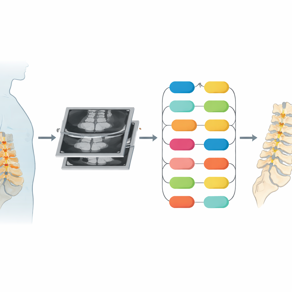
A Network That Pays Attention to Edges
The authors propose a new deep learning model, called BS‑Net (Boundary Sensitive‑Net), that is trained to be especially attentive to the borders of each vertebra. Built on a popular image‑analysis backbone, the system adds two key components. The first is a multi‑task edge processing module, which learns to recognize both the interior of each bone and its outline at the same time, using an attention mechanism that lets information about shape and edge quality reinforce one another. The second is a contextual bilateral fusion module, which fuses fine‑detail edge cues from early layers of the network with broader, high‑level information from deeper layers. Together with a special loss function that explicitly penalizes boundary mistakes, these modules teach BS‑Net to trace the bony margins sharply, even when they are irregular or poorly contrasted.
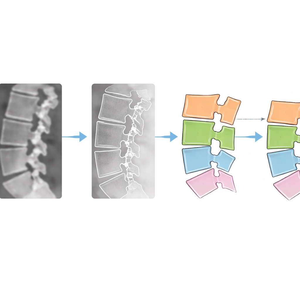
From Clean Outlines to Concrete Numbers
Once BS‑Net has separated each vertebra in a CT image, the system moves from pictures to measurements. It refines the outlines using a probabilistic smoothing step, then applies geometric techniques to locate each vertebra’s center and front‑to‑back edges. A minimum bounding rectangle method proved most accurate for finding the centers. With these landmarks, the system computes the angles between neighboring vertebrae and how far one bone has slipped relative to the one below, expressed as a percentage of its depth—mirroring the Meyerding grading scale used in clinics. Tested on 783 CT images from 379 patients and on an external MRI dataset, BS‑Net outperformed ten established segmentation models, reaching very high overlap scores between its automatic masks and expert‑drawn ones. Importantly, its slippage measurements agreed closely with manual assessments, with correlation values above 0.9 and smaller distance errors than competing methods.
What This Means for Patients and Clinics
In plain terms, the study shows that a spine‑focused AI can draw more precise outlines of the lower back bones and turn those outlines into trustworthy measurements of how much a vertebra has slipped. This could help radiologists and surgeons spot problematic slippage more consistently, track disease progression, and plan treatments with greater confidence, all while saving time. The authors caution that the method still struggles with very unusual cases, such as patients with metal implants or extreme deformities, and further training on diverse data will be needed. Even so, BS‑Net represents a significant step toward fully automated, quantitative assessment of lumbar spondylolisthesis from routine CT and MRI scans.
Citation: Ji, D., Qian, F. & Zong, Y. Boundary sensitive-net-based lumbar vertebra segmentation and spondylolisthesis measurement. Sci Rep 16, 13612 (2026). https://doi.org/10.1038/s41598-026-38522-7
Keywords: lumbar spine, spondylolisthesis, medical image segmentation, deep learning, vertebra CT analysis