Clear Sky Science · en
Development of a cell-subculture bell (C-Bell) device using low-cost RGB color sensors
Why watching cell dishes is harder than it sounds
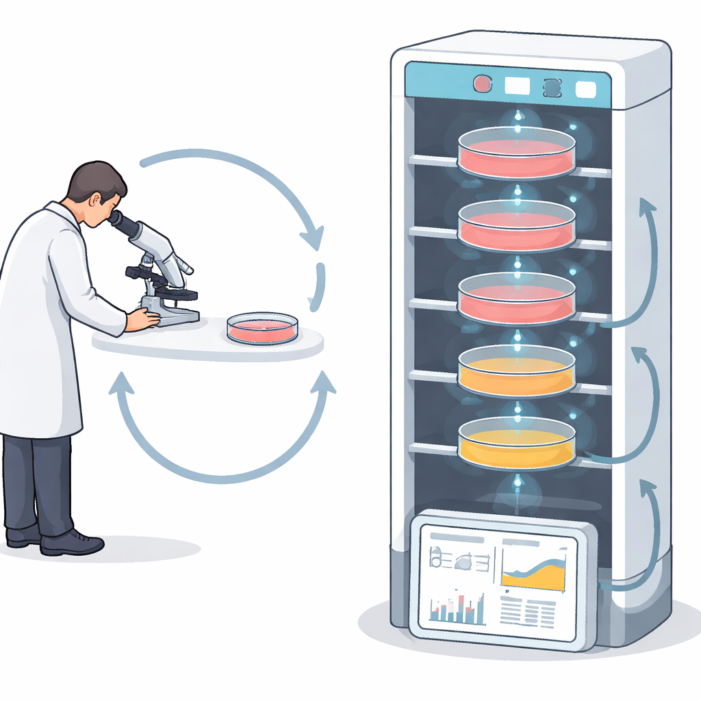
Much of modern biology and drug development depends on keeping living cells happy in plastic dishes. Researchers must regularly “split” or subculture these cells before they become overcrowded and stressed. Today, that timing is usually judged by eye: scientists peer through microscopes and glance at the color of the liquid that bathes the cells. This routine is time‑consuming, subjective, and easy to misjudge. The study introduces a simple, low‑cost device called C‑Bell that automatically watches over cell dishes by reading subtle color changes in the growth liquid, turning a mundane chore into an objective, hands‑off process.
A small helper built from everyday parts
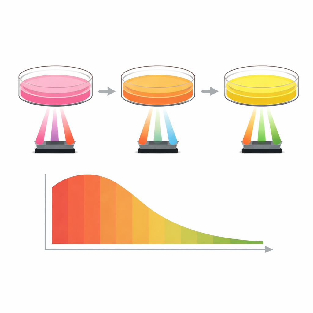
The C‑Bell device is built around widely available hobby electronics, including an Arduino microcontroller and an inexpensive RGB color sensor. Cells are grown in standard 60 mm plastic dishes filled with nutrient liquid that contains Phenol Red, a dye that shifts from red to yellow as the liquid becomes more acidic. As cells grow and burn fuel, they release acidic by‑products that slowly nudge the color toward yellow. C‑Bell sits underneath stacked dishes inside a regular carbon‑dioxide incubator and shines light through the bottom of each dish. The sensor measures how much red, green, and blue light is reflected. Because the device is self‑contained and battery‑powered, it does not require modifying the incubator or using special cultureware.
Turning color into a simple number
To make sense of the color readings, the researchers created a single scale called the C‑Bell Index. The device repeatedly samples the red, green, and blue signals and uses the green component, which changes most clearly as the liquid moves from red to yellow. After normalizing the data to remove sensor‑to‑sensor differences and compressing it into a 0–100 range, the team defined a practical threshold value. When the index stays high, the liquid is closer to its original pink color, and the cells still have room to grow. As the index falls below about 50, the liquid has become more yellow, signaling that the dish is crowded, metabolism is high, and the cells should be subcultured. This numeric readout replaces the researcher’s subjective color judgment with a reproducible, easy‑to‑track signal.
Putting the device to the test
First, the team confirmed that the color sensor could reliably distinguish between fresh, red culture medium and an experimentally acidified yellow version. The green light readings rose sharply in the yellow medium, and the calculated C‑Bell Index dropped from roughly the 80s down to around 20, matching direct pH measurements that showed a shift from about 7.4 to 6.7. Next, they tested C‑Bell with live lung cancer cells (A549), seeding dishes at different starting densities and following them over several days. By periodically moving dishes onto C‑Bell, or by leaving the device inside the incubator for fully automated hourly measurements, they tracked how quickly the index declined in each case. Dishes that started with many cells showed a rapid drop in the index and a fast change in liquid color from pink to yellow, while sparsely seeded dishes changed more slowly and never crossed the alarm threshold within the same time window.
Linking numbers, colors, and real cells
To ensure that the index really reflected what was happening in the dishes, the researchers compared C‑Bell readings with microscope images of the cells at key time points. When the index stayed above 60, images showed scattered cells and plenty of empty space, and the liquid remained pinkish. When the index hovered near 50, the cell layer was nearly continuous, and the liquid had shifted to an orange tone—an ideal moment to subculture. Once the index dropped well below 50, the liquid turned strongly yellow and the dishes were packed with cells, a condition associated with greater metabolic stress. Across repeated experiments, the device produced consistent index values with low variability, suggesting it could be trusted for day‑to‑day monitoring under the tested conditions.
Room to grow and future improvements
While promising, the current C‑Bell system has clear boundaries. It was tested with only one lung cancer cell line in a single type of growth medium that contains Phenol Red. Media without such a dye may not show the same visible color shift, limiting the approach unless alternative color indicators are used. The optical setup, based on a broad white LED and a low‑cost sensor, also leaves room for improvement in sensitivity and spectral matching, and the multi‑layer tower design has yet to be stressed in true high‑throughput scenarios. The authors propose future versions with better matched light sources, wireless data links, and validation across many cell types and media recipes.
What this means for everyday lab work
For non‑specialists, the main message is straightforward: C‑Bell turns the familiar “pink‑to‑yellow” color change in cell dishes into a continuous, objective clock that tells researchers when to take action. By using cheap electronics and simple optics, it offers small labs and early‑stage projects an accessible way to automate one of the most tedious parts of cell culture. If further refined and broadly validated, such devices could reduce human error, free scientists from constant dish‑watching, and make cell‑based experiments more reliable and comparable from one lab to another.
Citation: Koo, IS., Chang, S.J., Park, N.M. et al. Development of a cell-subculture bell (C-Bell) device using low-cost RGB color sensors. Sci Rep 16, 12130 (2026). https://doi.org/10.1038/s41598-026-38353-6
Keywords: cell culture monitoring, automated subculture, color sensor, low-cost biosensing, Arduino device