Clear Sky Science · en
Biointegration of a partially decellularized tracheal scaffold in a porcine model - preliminary results
New hope for children with damaged windpipes
Severe diseases of the windpipe, or trachea, can leave children with no good treatment options. When large sections of the airway are damaged, surgeons currently have no reliable replacement, and many young patients eventually die from their condition. This study explores a promising alternative: a specially prepared pig windpipe scaffold that might one day be used to rebuild children’s airways without lifelong anti‑rejection drugs.
Building a natural scaffold from a donor windpipe
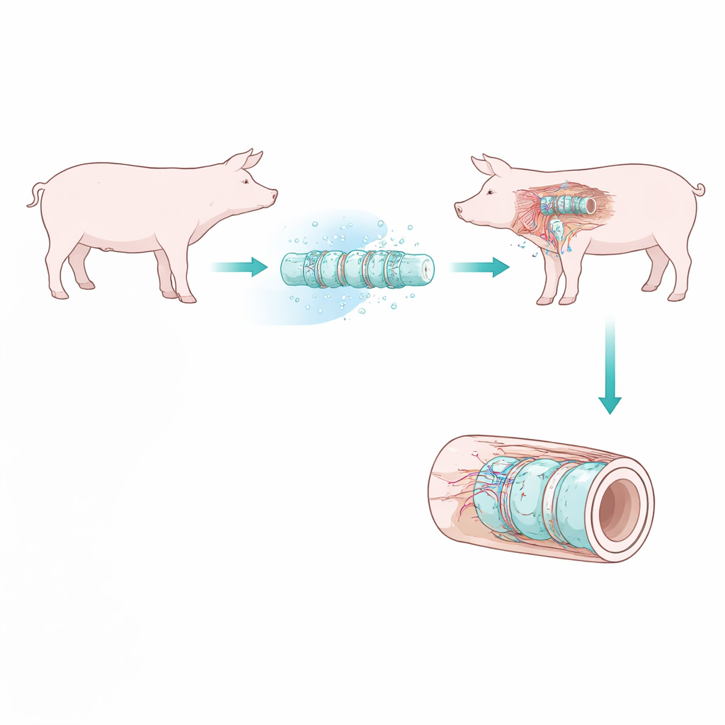
The researchers started from a simple idea: instead of using plastic tubes or full organ transplants, they would turn a donor trachea into a “shell” that the patient’s own body could inhabit. To do this, they partially removed the original pig cells with a series of washing steps, keeping the tough cartilage rings that hold the airway open while stripping away the inner lining and soft tissue that most strongly trigger immune attack. This process, called partial decellularization, leaves behind a clean, natural framework that is less likely to be rejected yet still mechanically sturdy enough to resist collapse during breathing.
Testing the scaffold inside living muscle
Before such a scaffold could replace a child’s airway, it must first connect to the body’s blood supply and be colonized by new cells. The team therefore implanted 11 of these prepared tracheas into the neck muscles of pigs, not as working airways but as a safe “training ground” where the graft could mature. Some pigs received the immune‑suppressing drug cyclosporin A, commonly given to transplant patients, while others did not. The tracheal pieces were left in place for either 28 days or 56 days before removal and detailed analysis, allowing the scientists to track how well the body accepted and remodeled the graft over time.
How the body welcomed the new tissue
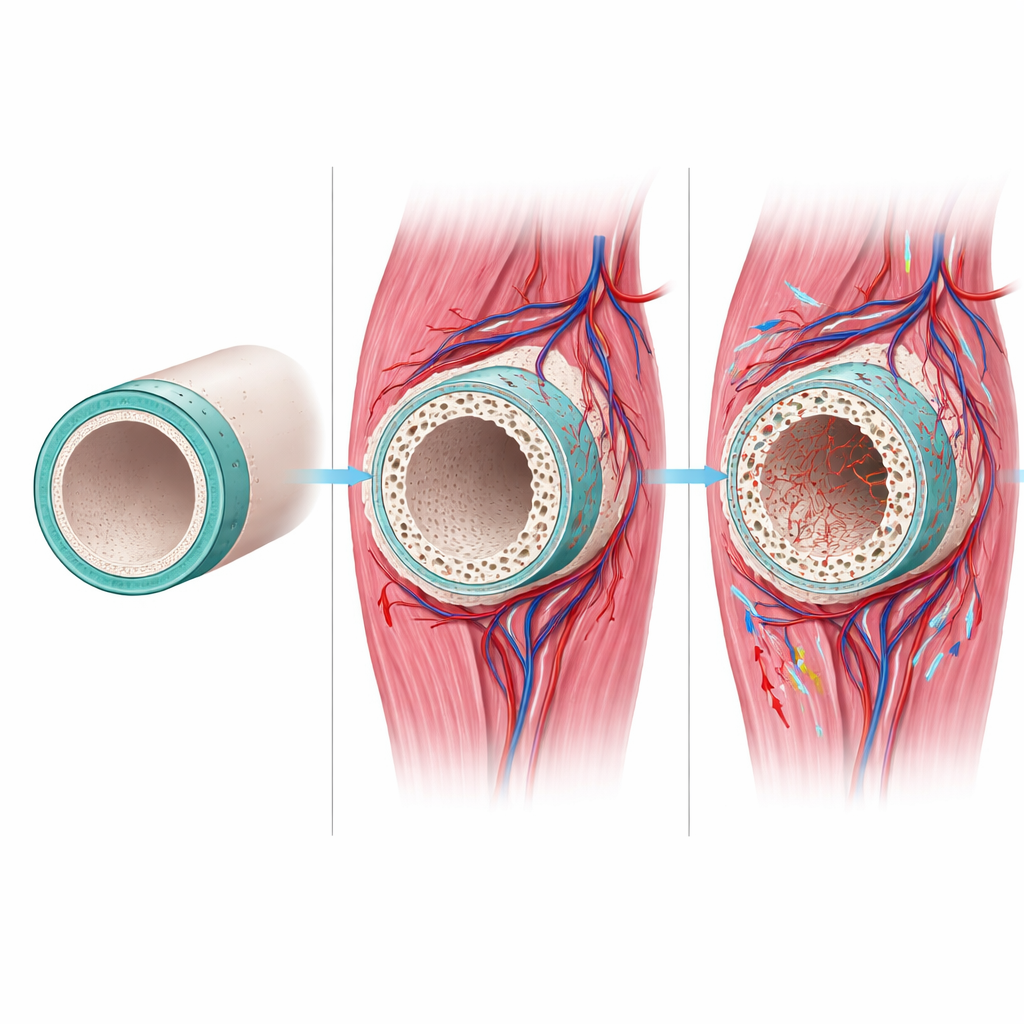
The implanted scaffolds behaved remarkably well. None of the pigs developed serious infections or signs of general illness, and blood tests showed no sustained inflammation. Under the microscope, the grafts were surrounded by healthy connective tissue rich in new blood vessels, and were being slowly colonized by fibroblasts—repair cells that lay down fresh supporting matrix. Immune cells that might signal rejection were present only in small numbers, similar to those seen in normal trachea. Importantly, these encouraging patterns were the same whether or not animals received cyclosporin A, suggesting that the partially cleaned scaffold was already “quiet” enough for the immune system to tolerate.
Striking a balance between strength and remodeling
The key structural concern was the cartilage, which must remain strong to keep a future airway open. The scientists found that freezing and thawing the grafts introduced fine cracks in the cartilage rings, and that these fissures became more pronounced after 56 days in the body than after 28 days. Chemical stains showed early signs of cartilage degradation and occasional patches of mineral buildup, but mechanical compression tests revealed that overall stiffness and resistance to collapse were preserved, and in some cases even slightly increased. New blood vessels and repair cells grew mainly around and between the rings rather than destroying them, indicating a controlled remodeling process rather than runaway damage.
Do anti‑rejection drugs really help here?
One of the most practical questions was whether patients with such scaffolds would truly need long‑term immunosuppressive drugs, which carry serious side effects, especially in children. Across multiple measures—local inflammation, systemic immune markers, vessel growth, and cell colonization—the study found no advantage to cyclosporin A treatment. The drug levels achieved in pigs confirmed that they were taking the medication, but the partially decellularized grafts simply did not provoke the kind of aggressive immune response seen with traditional organ transplants.
What this means for future airway repair
To a non‑specialist, the main message is that a carefully prepared pig windpipe can be accepted by another pig’s body with minimal fuss, becoming vascularized and repopulated by host cells while keeping its ability to hold an airway open. The work suggests that a 28‑day “maturation” period inside muscle, without immunosuppressive drugs, is enough to achieve good integration while limiting cartilage damage. Although these are early, small‑scale results in animals, they mark an important step toward a realistic, living replacement for damaged tracheas in children—one that might avoid both synthetic hardware and the burden of lifelong anti‑rejection therapy.
Citation: Vigouroux, A., Bonnin, Y., Gendron, N. et al. Biointegration of a partially decellularized tracheal scaffold in a porcine model - preliminary results. Sci Rep 16, 10121 (2026). https://doi.org/10.1038/s41598-026-37823-1
Keywords: tracheal replacement, tissue engineering, decellularized scaffold, pediatric airway, porcine model