Clear Sky Science · en
ScaleMamba-YOLO: a multi-scale MambaYOLO for medical object detection
Sharper Computer Eyes for Medical Scans
Doctors increasingly rely on computers to spot tiny warning signs in brain scans, blood samples, and endoscopy images. But these digital helpers often miss very small trouble spots or get distracted by harmless tissue that looks similar to disease. This paper introduces ScaleMamba‑YOLO, a new image analysis system designed to see both tiny and large medical problems more clearly and to ignore confusing background details, with the goal of making computer‑aided diagnosis more reliable in real time.
The Challenge of Seeing Illness in Pictures
Medical images are far more complicated than everyday photos. A single scan can contain tiny calcium specks, medium‑sized blood cells, and large tumors, all with fuzzy boundaries that blend into surrounding healthy tissue. Traditional detection programs either focus on one typical object size or try to handle everything at once, often at great computing cost. As a result, they can miss subtle lesions or mistake normal anatomy for disease, which is unacceptable when decisions about surgery or treatment are on the line.
A New Way to Look at Medical Images
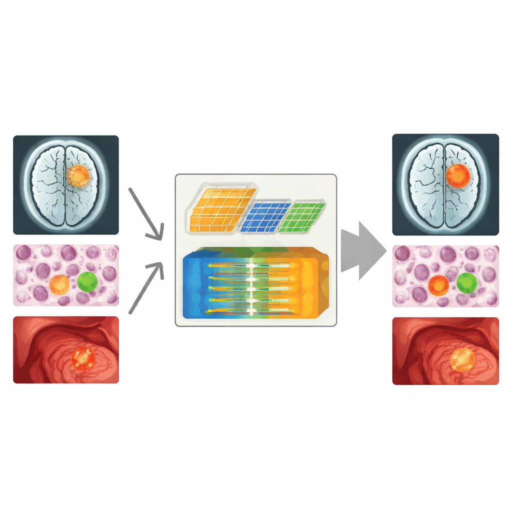
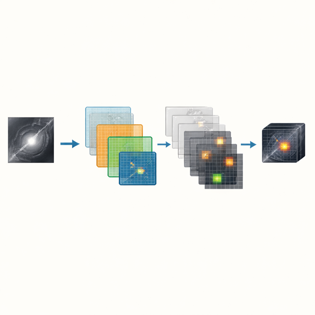
ScaleMamba‑YOLO builds on a fast object‑detection family known as YOLO and a newer “state‑space” engine called Mamba, which is good at understanding long‑range patterns. The authors carefully reshape this backbone for medical work by adding two key building blocks. First, a Medical Multi‑scale Local Feature Enhancement Block acts like three different camera lenses operating in parallel: one that zooms in on tiny details, one for mid‑range features, and one for broader regions. By combining these views, the system can spot both pinpoint lesions and broad, hazy abnormalities in a single pass.
Learning to Tune Out Background Noise
The second addition, called the Partial‑Enhanced C2f module, tackles a different problem: distracting background. In many scans, diseased and healthy tissues share similar shapes and shades, so a naïve model amplifies everything. Here, the authors introduce a selective operation that only performs heavy processing on a fraction of the information channels thought to be most important, while passing the rest through more lightly. This partial treatment acts like a smart filter, dampening features linked to normal tissue and sharpening those linked to possible lesions, without adding a large computing burden.
Proof from Brain, Blood, and Polyp Images
To test their design, the researchers ran ScaleMamba‑YOLO on three different medical image collections: brain MRI scans for tumor detection, microscope images of blood cells, and colonoscopy images of intestinal polyps. They also tried it on a standard everyday photo dataset to check whether the system overfits to medical pictures. Across all four sets, the new model consistently outperformed a strong earlier version (MambaYOLO) and several well‑known detectors, improving the main accuracy measure by roughly two percentage points on each dataset. It was especially strong at finding small objects such as tiny tumors, small blood cells, and hard‑to‑see polyps, while still running fast enough for real‑time video analysis on modern graphics cards.
What This Means for Future Care
For non‑specialists, the key message is that ScaleMamba‑YOLO makes computer vision tools better at spotting disease across a wide range of sizes while paying less attention to misleading background details. The method blends multi‑scale “eyes” with selective noise filtering, leading to cleaner, more trustworthy detections without sacrificing speed. Although more testing is needed on other scan types and in real hospitals, this work shows how carefully tailored AI architectures can turn messy medical images into clearer guidance for clinicians, potentially catching dangerous changes earlier and reducing missed findings.
Citation: Qin, X., Qian, Q., Li, X. et al. ScaleMamba-YOLO: a multi-scale MambaYOLO for medical object detection. Sci Rep 16, 10839 (2026). https://doi.org/10.1038/s41598-026-37258-8
Keywords: medical image analysis, lesion detection, deep learning, object detection, computer-aided diagnosis