Clear Sky Science · en
TREM1-PET imaging maps whole-body innate immune responses in a mouse model of metastatic melanoma
Why this matters for patients and families
When cancers spread to the brain, doctors often struggle to see how the immune system is responding deep inside the tumor and throughout the body. Standard brain scans show size and shape, but not whether immune cells are helping to fight the cancer or being turned off by it. This study tests a new type of whole-body scan in mice that can light up a specific group of immune cells linked to poor outcomes, offering a possible future tool to better predict how brain metastases will behave and respond to treatment.
A new way to see hidden immune activity
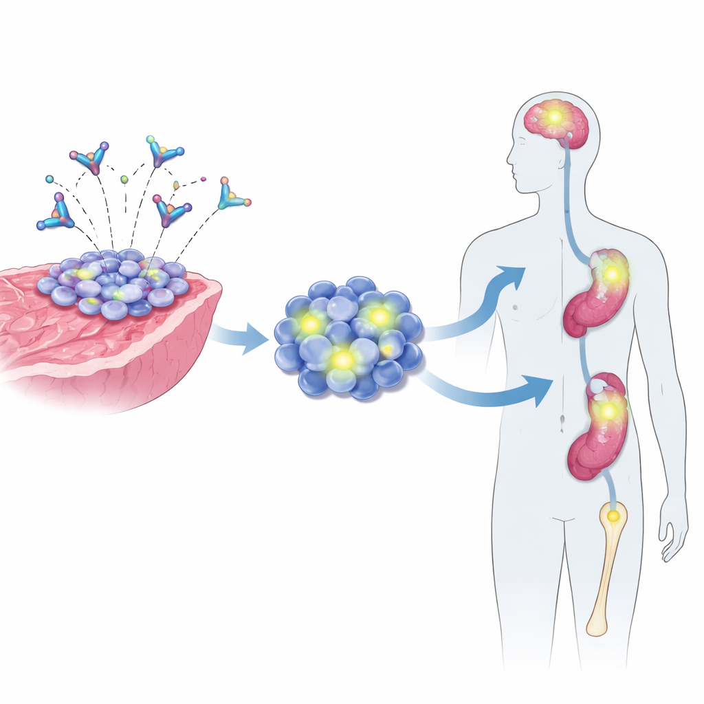
Many brain metastases blunt the body’s defenses by reshaping nearby immune cells, especially a group called tumor-associated myeloid cells. These cells can shut down the anti-tumor T cells that would otherwise attack the cancer. A surface receptor known as TREM1 is found at high levels on these myeloid cells in many cancers and is tied to tumor spread and shorter survival. The researchers developed a radioactive antibody, called [64Cu]TREM1-mAb, that homes in on TREM1. Attached to a PET scanner, this tracer has the potential to create a live, three-dimensional map of these immune cells in both the brain tumor and the rest of the body.
Putting the tracer to the test in brain cancer
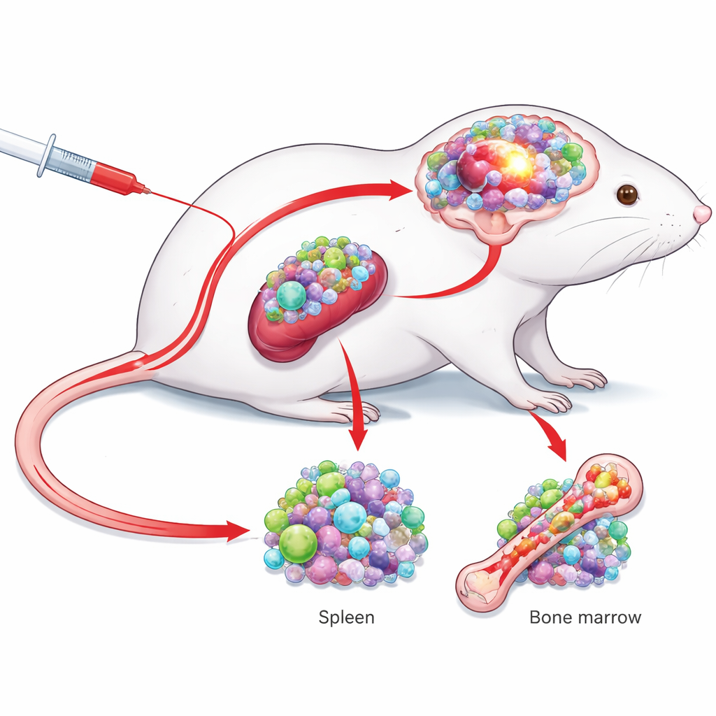
To see if this approach works in practice, the team implanted melanoma cells into the brains of mice to mimic metastatic brain tumors, and carried out sham brain surgeries in control animals. They injected the TREM1-targeted tracer or a non-targeted “isotype” tracer into the animals’ veins and imaged them with PET and MRI at two time points. By 48 hours after injection, the TREM1 tracer produced a much stronger signal in the tumor-containing side of the brain than in the opposite side, and much higher than in sham-operated brains. When the same analysis was done using the non-targeted tracer, this difference largely disappeared, showing that the bright signal in the tumors depended on TREM1 binding rather than just leaky blood vessels or surgical damage.
Seeing the whole-body immune response
Brain tumors can influence the immune system far beyond the skull. The investigators therefore examined classic immune organs such as bone marrow and spleen. PET images showed higher TREM1 tracer uptake in the bone marrow and, after correcting for blood levels, clearly higher standardized uptake ratios in both bone marrow and spleen of tumor-bearing mice compared with sham or isotype-tracer controls. Follow-up measurements in dissected tissues confirmed that more of the TREM1 tracer had actually accumulated in blood, bone marrow, muscle, and spleen in tumor-bearing animals. These patterns suggest that the brain tumor was driving a broader, whole-body activation of TREM1-positive myeloid cells, not just a local change at the tumor site.
Zooming in on which cells are lighting up
To identify the exact cells responsible for the signal, the team used several high-resolution methods. Autoradiography of thin brain slices showed that the tracer concentrated precisely within the tumor area, matching standard tissue stains. Flow cytometry, a technique that profiles individual cells, revealed that TREM1 was strongly expressed on a specific population of brain and spleen myeloid cells, but not on resident brain support cells or on T and B lymphocytes. Additional analysis of human single-cell RNA data from patients with brain metastases showed a similar pattern: TREM1 was present in certain myeloid cells but not in cancer cells or most other immune cells. Together, these findings tie the PET signal tightly to a defined, cancer-associated myeloid population.
What this could mean for future care
By showing that a TREM1-targeted PET tracer can detect harmful immune activity both in brain tumors and in distant immune organs in mice, this work lays the groundwork for a new kind of imaging in patients with brain metastases. Unlike current markers that light up many different cell types, TREM1 focuses attention on myeloid cells that are known to aid tumor growth and predict worse outcomes. If human versions of this tracer can be developed and optimized, clinicians could one day use TREM1-PET scans to monitor how aggressively the tumor is subverting the immune system, track whether immunotherapies are reawakening anti-tumor responses, and tailor treatment plans more precisely, potentially improving survival and quality of life.
Citation: Falk, I.N., Chaney, A.M., Verma, R. et al. TREM1-PET imaging maps whole-body innate immune responses in a mouse model of metastatic melanoma. Sci Rep 16, 11157 (2026). https://doi.org/10.1038/s41598-026-36542-x
Keywords: brain metastases, immune imaging, PET tracer, myeloid cells, melanoma