Clear Sky Science · en
An open-access multi-site fMRI dataset for investigating conscious visual perception
Why this matters to everyday seeing
Every moment, your eyes send a flood of images to your brain, yet only some of them reach conscious awareness. This article introduces a large, openly shared brain imaging dataset that lets scientists probe how clearly visible pictures of faces, objects, and letters are processed when we pay attention to them or ignore them. By making these detailed recordings freely available, the project invites researchers worldwide to tackle one of science’s biggest puzzles: how brain activity gives rise to what we actually see.
Looking inside the awake visual brain
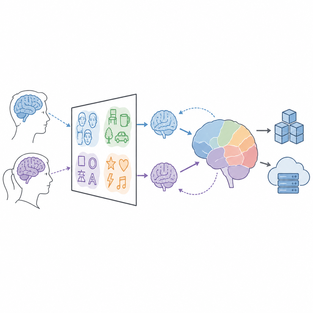
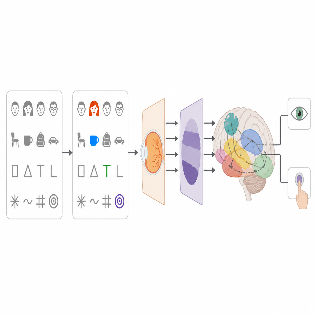
The researchers used functional magnetic resonance imaging (fMRI) to measure brain activity in 118 healthy adults while they viewed simple, easily seen images. On each trial, people saw one picture in the center of a screen: a face, an everyday object, a letter, or a made up “false font,” each shown from different viewpoints and for three viewing times between half a second and one and a half seconds. Because the pictures were clear and unambiguous, any failures of a theory of consciousness to match the data cannot be blamed on weak or borderline perception, making this a strong testbed for ideas about conscious vision.
Testing rival ideas about awareness
The dataset was created within the Cogitate consortium, a large adversarial collaboration that brings together supporters of two leading theories of consciousness: Global Neuronal Workspace theory and Integrated Information Theory. Instead of each camp collecting its own data and arguing past the other, both sides agreed on a shared experimental design in advance and on how the brain signals would be analyzed. In the task, only a few pictures in each block were designated as targets that required a button press. This split the constant stream of clearly visible images into task relevant and task irrelevant groups, allowing scientists to ask how attention and goal directed behavior shape what becomes part of our conscious experience.
A simple task with rich possibilities
Although the experiment itself was straightforward, it was carefully structured to produce many lines of evidence from the same data. The type of picture, its orientation, its duration on the screen, and its relevance for the task were all varied in a systematic way. Participants also had their gaze tracked, so researchers can confirm that people looked where they were supposed to, and their moment by moment behavior was recorded. Because the same protocol was run at two independent imaging centers with matching scanners and eye trackers, the dataset allows tests of whether findings hold across different sites and equipment, a key step toward robust, reproducible science.
How the data are organized for sharing
To maximize re use, the team converted the anonymized scans and auxiliary files into a widely adopted standard known as the Brain Imaging Data Structure. Each volunteer’s folder contains structural brain images, the fMRI recordings from the task, supporting scans used to correct image distortions, and detailed timing files describing exactly which picture appeared when, for how long, and with what task status. Additional files describe the equipment, experimental protocols, demographics, and quality checks. The same information can be downloaded either as zipped bundles or browsed through an online database that also provides an application programming interface for automated access.
From raw signals to insights about seeing
The authors carried out extensive quality control before releasing the data. They checked for missing files, stripped all personal identifiers, and inspected measures of motion, sharpness, and signal to noise in both structural and functional scans, excluding only two participants with clear artifacts. Eye movement records confirmed that most volunteers maintained good fixation, and behavioral measures showed very high accuracy in detecting the rare targets. Along with public code for preprocessing and analysis, these steps mean that other groups can confidently build on the resource to ask their own questions about visual processing, time perception, or the role of task relevance in shaping brain activity.
What this means for our understanding of vision
Instead of claiming to settle the debate about consciousness, this work provides a solid, transparent foundation on which many investigations can rest. By combining a clean experimental design, large sample size, careful quality checks, and open formats, the dataset lets researchers see how the brain responds to clearly visible images under changing task demands. For a lay reader, the key message is that progress on the mystery of conscious vision now depends less on isolated, one off experiments and more on shared resources like this one, which allow competing ideas to be tested and compared fairly using the same rich stream of brain data.
Citation: Khalaf, A., Richter, D., Vidal, Y. et al. An open-access multi-site fMRI dataset for investigating conscious visual perception. Sci Data 13, 779 (2026). https://doi.org/10.1038/s41597-026-07377-y
Keywords: consciousness, visual perception, fMRI dataset, brain imaging, attention