Clear Sky Science · en
A fundus image dataset for intelligent diabetic retinopathy system
Why this matters for people with diabetes
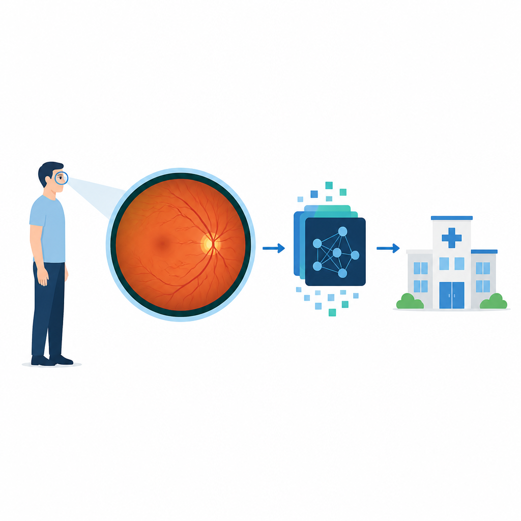
Diabetes can quietly damage the eyes long before vision starts to blur. This paper describes a new image collection designed to help computers spot these hidden eye changes earlier and more reliably, using a wider view of the retina than most current tools provide.
Seeing more of the back of the eye
Diabetic eye disease harms the tiny blood vessels at the back of the eye and is a leading cause of permanent vision loss in working age adults. Regular eye photos are already used to look for trouble, but they capture only a small central area. Important warning signs often appear at the edges of the retina, where traditional cameras may simply not see them. Ultra wide field imaging solves this by capturing as much as five times more area in a single shot, including the far corners where early damage and new, fragile blood vessels can arise.
Building a rich picture library for smart tools
To make the most of this wider view, computer programs need large, well organized sets of images to learn from. The authors collected 1,630 ultra wide field eye photos from 809 patients at two hospitals in China. All people in the study had diabetes, and their average age was about 54 years. The images were taken with a special camera that does not require eye drops to widen the pupil, making the test quicker and more comfortable. Low quality images with blur or missing areas were removed so that only clear views of the retina, blood vessels, and possible damage remained.
How experts taught the computer
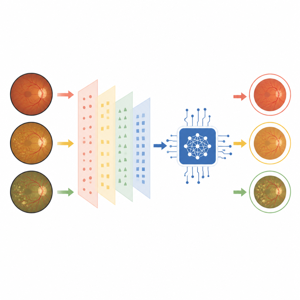
Three experienced eye doctors carefully examined every image and sorted them into three groups: normal eyes, eyes with early to moderate damage, and eyes with advanced damage. They followed international rules for grading diabetic eye disease, but the authors note that some disagreement between doctors is still expected, especially when deciding whether very mild changes should count as normal or early disease. The final decisions were recorded in folders and simple tables that list each image and its group, creating an easy starting point for other research teams.
Putting the dataset to the test
The team then checked how useful this image library is for training artificial intelligence. They split the images into training, validation, and test sets and ran four well known deep learning models to classify the three stages of disease. Before training, they cropped and standardized the pictures and used simple tricks such as random flips to help the models generalize. On the held out test images, all models achieved strong scores across several measures of accuracy, with one model in particular performing the best. Heatmaps from the models highlighted tiny bulges in blood vessels and scar like membranes, suggesting that the algorithms focused on the same areas that doctors consider important.
Helping more people keep their sight
This new open dataset offers a high quality, wide angle view of diabetic eye disease for anyone developing smart screening tools. By combining ultra wide field images with artificial intelligence, future systems could cover more of the retina, miss fewer dangerous changes, and require less staff time. While this dataset alone cannot replace regular eye care, it lays the groundwork for more reliable, widely available computer aided screening that could help reduce blindness from diabetes, especially in clinics with limited resources.
Citation: Peng, S., Yang, S., Zhao, X. et al. A fundus image dataset for intelligent diabetic retinopathy system. Sci Data 13, 777 (2026). https://doi.org/10.1038/s41597-026-07093-7
Keywords: diabetic retinopathy, retinal imaging, ultra wide field, medical AI, eye screening